A study on the relationship between clinical and CT features and length of stay of general type novel coronavirus pneumonia patients in mobile cabin hospital
-
摘要:
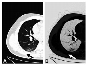
目的 回顾性分析新型冠状病毒肺炎普通型患者的临床和胸部CT特征及对患者住院时间的影响,为认识新冠肺炎的影像学特征、判断转归提供参考。 方法 总结武汉市东西湖方舱医院2020年2月7日—2020年3月3日收治的84例新型冠状病毒肺炎普通型患者的临床和胸部CT影像特点,使用回归分析判断患者临床和影像特点对住院时间的影响。 结果 全组84例患者中,男性41例,女性43例,年龄17~73岁,核酸复检阳性者15例(17.86%)。患者胸部CT影像学表现最常见有磨玻璃影(98.81%)和血管增粗影(89.29%),大部分患者出现血管增粗并与胸膜垂直表现(51.19%),部分患者可出现Kerley B线(47.62%)、条索影(38.10%)及实变影(16.67%),少部分患者可出现结节影(8.33%)和铺路石征(4.76%)。多数患者病变为双侧分布(82.14%),病变同时累及中心和周边区域的比例为11.90%,仅累及周边者88.10%。本组病例住院时间为8~25 d,平均住院时间为(16.02±4.32)d。患者核酸复检阳性者较阴性者住院时间延长4.179 d,出现病变同时累及中心和周边区域、血管增粗并与胸膜垂直征象者较无上述影像特征者住院时间分别延长2.692 d和3.123 d。 结论 核酸复检阳性、病变累及中心区域、有血管增粗并与胸膜垂直征象者住院时间延长,应及时与患者沟通、做好心理疏导。 Abstract:Objective To retrospectively analyze the clinical and CT features of novel coronavirus pneumonia patients with general type and its' influence on the length of hospital stay, so as to provide reference for understanding the imaging features of novel coronavirus pneumonia and determining the outcome of patient. Methods The clinical and CT features of 84 patients with general type novel coronavirus pneumonia admitted to Wuhan Dongxihu mobile cabin hospital from February 7, 2020 to March 3, 2020 were summarized. Regression analysis was used to determine the impact of clinical and imaging characteristics on hospital stay. Results Among the 84 patients, 41 were males and 43 were females, aged from 17 to 73 years old, and 15 cases were positive for nucleic acid reexamination (17.86%). The most common manifestations of chest CT imaging were ground glass (98.81%) and enlarged vascular lumens (89.29%%). Most of the patients had signs of enlarged vascular lumens perpendicular to pleura (51.19%), some patients had Kerley B line (47.62%), strip shadow (38.10%) and consolidation shadow (16.67%), and a few patients had nodules (8.33%) and paving stone sign (4.76%). The majority of cases had bilateral distribution of lesions (82.14%), the proportion of lesions involving both the central and peripheral regions was 11.90%, and only the peripheral areas was 88.10%. The length of hospital stay was 8-25 days, and the average length of hospital stay was (16.02±4.32) days. The hospital stay of patients with positive nucleic acid reexamination was extended by 4.179 days compared with those with negative nucleic acid retest. The hospital stay of patients with lesions involving both central area and peripheral area, enlarged vascular lumens perpendicular to pleura was extended by 2.692 days and 3.123 days respectively compared with those without the above image features. Conclusion The length of hospital stay was longer in patients with positive nucleic acid reexamination, involvement of central area, thickening of blood vessels and signs perpendicular to pleura. It should be communicate with patients in time, and do a good job in psychological counseling. -
Key words:
- Mobile cabin hospital /
- SARS-CoV-2 /
- COVID-19 /
- Clinical characteristics /
- Computer Tomography /
- Imaging features /
- Hospital stays
-
表 1 临床特点及胸部CT表现数据分布
项目 类别 病例数(%) 性别 女性 43(51.19) 男性 41(48.81) 年龄 <60岁 65(77.38) ≥60岁 19(22.62) 核酸结果 阴性 69(82.14) 阳性 15(17.86) 同时累及中心和周边区域 无 74(88.10) 有 10(11.90) 双侧分布 否 15(17.86) 是 69(82.14) 磨玻璃 无 1(1.19) 有 83(98.81) 实变影 无 70(83.33) 有 14(16.67) 结节影 无 77(91.67) 有 7(8.33) 条索影 无 52(61.90) 有 32(38.10) 空气支气管征 无 83(98.81) 有 1(1.19) 机化性肺炎 无 70(83.33) 有 14(16.67) 血管增粗影 无 9(10.71) 有 75(89.29) 血管增粗并与胸膜垂直 无 41(48.81) 有 43(51.19) Kerley B线 无 44(52.38) 有 40(47.62) 铺路石征 无 80(95.24) 有 4(4.76) 表 2 医嘱离院患者住院时间影响因素的相关性分析结果
相关系数 P值 性别 0.144 0.190 年龄分组 0.077 0.489 核酸结果 0.454 < 0.001 同时累及中心和周边区域 0.315 0.004 磨玻璃影 0.001 0.996 实变影 0.146 0.184 结节影 0.048 0.661 条索影 0.241 0.027 空气支气管征 0.025 0.822 机化性肺炎 0.146 0.184 血管增粗影 0.253 0.020 血管增粗并与胸膜垂直 0.521 <0.001 Kerley B线 0.400 < 0.001 铺路石征 -0.118 0.283 双侧分布 0.111 0.314 表 3 住院时间影响因素的线性回归分析结果
变量 B SE β t值 P值 95%CI 核酸结果 4.179 0.976 0.373 4.281 < 0.001 2.235~6.122 同时累及中心和周边区域 2.692 1.225 0.203 2.198 0.031 0.253~5.130 条索影 0.425 0.852 0.048 0.499 0.619 -1.271~2.121 血管增粗影 0.928 1.267 0.067 0.732 0.466 -1.596~3.451 血管增粗并与胸膜垂直 3.123 1.134 0.364 2.753 0.007 0.864~5.382 Kerley B线 -0.019 1.088 -0.002 -0.018 0.986 -2.185~2.147 截距 12.377 1.119 11.066 < 0.001 10.150~14.605 -
[1] CHEN S M, ZHANG Z J, YANG J T, et al. Fangcang shelter hospitals: a novel concept for responding to public health emergencies[J]. Lancet, 2020, 395(10232): 1305-1314. doi: 10.1016/S0140-6736(20)30744-3 [2] 佚名. 新型冠状病毒感染的肺炎诊疗方案(试行第五版)[J]. 江苏中医药, 2020, 52(2): 95-96. https://www.cnki.com.cn/Article/CJFDTOTAL-JSZY202002034.htm [3] 中华医学会放射学分会. 新型冠状病毒肺炎的放射学诊断: 中华医学会放射学分会专家推荐意见(第一版)[J]. 中华放射学杂志, 2020, 54(4): 279-285. doi: 10.3760/cma.j.cn112149-20200205-00094 [4] 钟飞扬, 张寒菲, 王彬宸, 等. 新型冠状病毒肺炎的CT影像学表现[J]. 武汉大学学报(医学版), 2020, 41(3): 345-348. https://www.cnki.com.cn/Article/CJFDTOTAL-HBYK202003001.htm [5] KANNE J P. Chest CTfindings in 2019 novel coronavirus (2019-nCoV) infections from Wuhan, China: Key points for the radiologist[J]. Radiology, 2020, 295(1): 16-17. doi: 10.1148/radiol.2020200241 [6] DAS K M, LEE E Y, LANGER R D, et al. Middle east respiratory syndrome coronavirus: What does a radiologist need to know?[J]. Am J Roentgenol, 2016, 206(6): 1193-1201. doi: 10.2214/AJR.15.15363 [7] CHOJ W J, LEE K N, KANG E J, et al. Middle east respiratory syndrome-coronavirus infection: A case report of serial computed tomographic findings in a young male patient[J]. Korean J Radiol, 2016, 17(1): 166-170. doi: 10.3348/kjr.2016.17.1.166 [8] HUANG C L, WANG Y M, LI X W, et al. Clinical features of patients infected with 2019 novel coronavirus in Wuhan, China[J]. Lancet, 2020, 395(10223): 497-506. doi: 10.1016/S0140-6736(20)30183-5 [9] 唐光孝, 李春华, 刘雪艳, 等. 新型冠状病毒肺炎的临床与CT表现[J]. 中国呼吸与危重监护杂志, 2020, 19(2): 161-165. https://www.cnki.com.cn/Article/CJFDTOTAL-ZGHW202002024.htm [10] 吴婧, 冯连彩, 冼新源, 等. 新型冠状病毒肺炎130例CT分布特点及征象研究[J]. 中华结核和呼吸杂志, 2020, 43(4): 321-326. [11] 高璐, 张静平, 杜永浩, 等. 输入性新型冠状病毒肺炎的CT表现. 西安交通大学学报(医学版)[J]. 西安交通大学学报(医学版), 2020, 41(3): 429-434. [12] 陈甜, 蒋宗焰, 许炜, 等. 76例新型冠状病毒肺炎患者的临床及CT影像特征分析[J]. 暨南大学学报(自然科学与医学版), 2020, 41(2): 157-162. https://www.cnki.com.cn/Article/CJFDTOTAL-JNDX202002010.htm [13] 曹佳, 周军, 廖星男, 等. 老年新型冠状病毒肺炎患者的临床特点与CT征象[J]. 武汉大学学报(医学版), 2020, 41(4): 551-554. https://www.cnki.com.cn/Article/CJFDTOTAL-HBYK202004008.htm [14] 刘茜, 王荣帅, 屈国强, 等. 新型冠状病毒肺炎死亡尸体系统解剖大体观察报告[J]. 法医学杂志, 2020, 36(1): 21-23. https://www.cnki.com.cn/Article/CJFDTOTAL-FYXZ202001006.htm [15] MOHANTY S K, SATAPATHY A, NAIDU M M, et al. Severe acute respiratory syndrome coronavirus-2 (SARS-CoV-2) and coronavirus disease 19 (COVID-19)-anatomic pathology perspective on current knowledge[J]. Diagn Pathol, 2020, 15(1): 727-733. http://www.ncbi.nlm.nih.gov/pubmed/32799894 -


 下载:
下载:


