Relationship between Livin expression and epithelial-mesenchymal transformation in oral squamous cell carcinoma and its clinical significance
-
摘要:
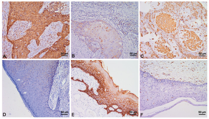
目的 探讨Livin蛋白和上皮间质转化(EMT)相关蛋白Vimentin、E-cadherin在口腔鳞状细胞癌(OSCC)组织中的表达,探讨三者表达的相关性及其临床意义。 方法 应用免疫组化EliVision法检测56例OSCC组织及56例癌旁组织中Livin、Vimentin、E-cadherin的表达,并分析三者表达的相关性及其与OSCC患者临床病理特征的关系。进一步应用Western blotting检测10例新鲜OSCC组织中Livin、Vimentin、E-cadherin蛋白的表达水平。 结果 Livin在OSCC组织中的阳性率为67.9%,明显高于癌旁组织中Livin的阳性率(16.1%),且Livin表达与OSCC的淋巴结转移及TNM分期相关。E-cadherin、Vimentin在OSCC组织中的阳性率分别为44.6%、50.0%,Livin与E-cadherin表达呈负相关,与Vimentin表达呈正相关。Western blotting检测结果显示Livin高表达的OSCC组织中,E-cadherin呈低表达,Vimentin呈高表达。 结论 Livin表达可促进OSCC侵袭转移,这种促进作用可能是通过表达Livin从而激活上皮间质转化(EMT)发生实现的。 Abstract:Objective To investigate the expression of Livin protein and epithelial-mesenchymal transformation (EMT)-related proteins Vimentin and E-cadherin in oral squamous cell carcinoma (OSCC) tissues, and to explore the correlation and clinical significance of the three protein expressions. Methods EliVision method of immunohistochemical was used to examine the expression of Livin, Vimentin and E-cadherin in 56 cases of OSCC and 56 cases of paracancerous tissues. The correlation between the expression of Livin, Vimentin and E-cadherin and the clinicopathological features of OSCC patients was analyzed. Furthermore, western blot was used to examine the expression of Livin, Vimentin and E-cadherin in 10 fresh OSCC tissues. Results The positive rate of Livin in OSCC tissues was 67.9%, which was significantly higher than that in paracancerous tissues (16.1%), and the expression of Livin was related to lymph node metastasis and TNM stage of OSCC. The positive rates of E-cadherin and Vimentin in OSCC tissues were 44.6% and 50%, respectively. Livin was negatively correlated with the expression of E-cadherin and positively correlated with the expression of Vimentin. The results of western blot showed that in the OSCC tissues with high expression of Livin, the expression of E-cadherin was low and Vimentin was high. Conclusion The expression of Livin can promote the invasion and metastasis of OSCC, which may be achieved by activating the occurrence of EMT by expressing Livin. -
Key words:
- Oral squamous cell carcinoma /
- Livin protein /
- Epithelial-mesenchymal transition /
- Invasion /
- Metastasis
-
表 1 Livin、E-cadherin、Vimentin在口腔鳞癌组织及癌旁组织中的表达
[个(%)] 组织类别 标本数 Livin E-cadherin Vimentin 肿瘤组织 56 38(67.9) 25(44.6) 28(50.0) 癌旁组织 56 9(16.1) 51(91.1) 5(8.9) χ2值 30.832 27.673 22.727 P值 < 0.001 < 0.001 < 0.001 表 2 Livin、E-cadherin、Vimentin表达与OSCC临床病理特征的关系
(例) 临床病理参数 例数 Livin表达 E-cadherin表达 Vimentin表达 + - χ2值 P值 + - χ2值 P值 + - χ2值 P值 年龄(岁) 0.983 0.322 0.024 0.877 0.292 0.589 ≤65 32 20 12 14 18 15 17 >65 24 18 6 11 13 13 11 性别 0.017 0.896 < 0.001 0.984 2.947 0.086 男性 38 26 12 17 21 16 22 女性 18 12 6 8 10 12 6 部位 0.721 0.949 1.864 0.761 4.486 0.344 舌 20 14 6 9 11 9 11 颊 14 10 4 6 8 8 6 唇 12 7 5 5 7 7 5 口底 6 4 2 2 4 1 5 牙龈 4 3 1 3 1 3 1 临床分期 4.912 0.027 5.901 0.015 6.171 0.013 Ⅰ+Ⅱ 35 20 15 20 15 13 22 Ⅲ+Ⅳ 21 18 3 5 16 15 6 分化程度 2.913 0.088 5.396 0.020 16.047 < 0.001 高+中 38 23 15 21 17 12 26 低+未 18 15 3 4 14 16 2 淋巴结转移 8.828 0.003 7.042 0.008 14.674 < 0.001 无 34 18 16 20 14 10 24 有 22 20 2 5 17 18 4 表 3 OSCC中Livin表达与EMT相关标志蛋白E-cadherin、Vimentin表达之间的关系
(个) EMT标志物 样本数 Livin r值 P值 + - E-cadherin + 25 10 15 -0.536 < 0.001 - 31 28 3 Vimentin + 28 24 4 0.382 0.004 - 28 14 14 -
[1] SU Q B, WANG L Y, WEI G N, et al. Livin serves as a prognostic marker for mid-distal rectal cancer and a target of mid-distal rectal cancer treatment[J]. Oncol Lett, 2017, 14(6): 7759-7766. http://www.onacademic.com/detail/journal_1000040115902010_b674.html [2] LIU A H, HE A B, TONG W X, et al. Prognostic significance of Livin expression in nasopharyngeal carcinoma after radiotherapy[J]. Cancer Radiother, 2016, 20(5): 384-390. doi: 10.1016/j.canrad.2016.05.013 [3] GU J, REN L, WANG X, et al. Expression of livin, survivin and caspase-3 in prostatic cancer and their clinical significance[J]. Int J Clin Exp Pathol, 2015, 8(11): 14034-14039. http://europepmc.org/articles/PMC4713502/pdf/ijcep0008-14034.pdf [4] XU H, CHENG Z, LIU D W, et al. High expression of Livin serves as a predictive indicator for parotid gland tumors[J]. Eur Rev Med Pharmacol Sci, 2019, 23(12): 5223-5228. http://www.ncbi.nlm.nih.gov/pubmed/31298372 [5] DUAN W J, Bi P D, MA Y, et al. MiR-512-3p regulates malignant tumor behavior and multi-drug resistance in breast cancer cells via targeting Livin[J]. Neoplasma, 2020, 67(1): 102-110. doi: 10.4149/neo_2019_190106N18 [6] GE Y, LIU B L, CUI J P, et al. Livin promotes colon cancer progression by regulation of H2A. XY39ph via JMJD6[J]. Life Sci, 2019, 234: 116788. doi: 10.1016/j.lfs.2019.116788 [7] HAN Y, ZHANG L, WANG W, et al. Livin promotes the progression and metastasis of breast cancer through the regulation of epithelial mesenchymal transition via the p38/GSK3β pathway[J]. Oncol Rep, 2017, 38(6): 3574-3582. http://europepmc.org/abstract/MED/29039608 [8] GE Y, CAO X, WANG D, et al. Overexpression of Livin promotes migration and invasion of colorectal cancer cells by induction of epithelial-mesenchymal transition via NF-κB activation[J]. Onco Targets Ther, 2016, 9: 1011-1021. http://pubmedcentralcanada.ca/pmcc/articles/PMC4778785/ [9] LIU S, LI X, LI Q, et al. Silencing Livin improved the sensitivity of colon cancer cells to 5-fluorouracil by regulating crosstalk between apoptosis and autophagy[J]. Oncol Lett, 2018, 15(5): 7707-7715. http://www.researchgate.net/publication/323811346_Silencing_Livin_improved_the_sensitivity_of_colon_cancer_cells_to_5-fluorouracil_by_regulating_crosstalk_between_apoptosis_and_autophagy/fulltext/5aabfa7faca2721710f89d74/323811346_Silencing_Livin_improved_the_sensitivity_of_colon_cancer_cells_to_5-fluorouracil_by_regulating_crosstalk_between_apoptosis_and_autophagy.pdf?origin=publication_detail [10] DONGRE A, WEINBERG R A. New insights into the mechanisms of epithelial-mesenchymal transition and implications for cancer[J]. Nat Rev Mol Cell Biol, 2019, 20(2): 69-84. http://www.nature.com/articles/s41580-018-0080-4 [11] DIEPENBRUCK M, CHRISTOFORI G. Epithelial-mesenchymal transition(EMT) and metastasis: yes, no, maybe?[J]. Curr Opin Cell Biol, 2016, 43: 7-13. doi: 10.1016/j.ceb.2016.06.002 [12] MITTAL V. Epithelial Mesenchymal Transition in Tumor Metastasis[J]. Annu Rev Pathol, 2018, 13: 395-412. doi: 10.1146/annurev-pathol-020117-043854 [13] 韩艳春, 崔敏, 张骞, 等. Livin表达与胃癌上皮-间质转化的关系及临床意义[J]. 临床与实验病理学杂志, 2018, 34(11): 1200-1202. [14] 张骞, 韩艳春, 曹璋, 等. Livin在结肠癌中的表达及与上皮-间质转化的关系[J]. 中国组织化学与细胞化学杂志, 2018, 27(3): 242-246. [15] ZHOU J, JIANG H. Livin is involved in TGF-β1-induced renal tubular epithelial-mesenchymal transition through lncRNA-ATB[J]. Ann Transl Med, 2019, 7(18): 463. doi: 10.21037/atm.2019.08.29 [16] 张生军, 常琦, 刘勇峰, 等. Livin通过Akt信号途径促进胃癌SGC7901细胞的上皮细胞间质转换[J]. 基础医学与临床, 2016, 36(5): 586-589. [17] ZHUANG L, SHEN L D, LI K, et al. Inhibition of livin expression suppresses cell proliferation and enhances chemosensitivity to cisplatin in human lung adenocarcinoma cells[J]. Mol Med Rep, 2015, 12(1): 547-552. doi: 10.3892/mmr.2015.3372 -


 下载:
下载:


