

Expression and significance of IL-17 and IL-22 in tuberculosis heat-resistant antigen and phospho-antigen stimulated cell amplified polarization
-
摘要:
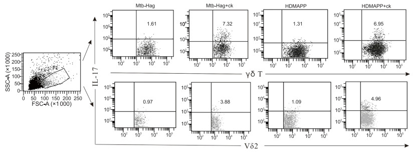
目的 γδ T细胞在结核耐热性抗原(Mtb-HAg)和磷酸化抗原(HDMAPP)培养扩增后,阐明Mtb-HAg和HDMAPP在细胞因子(ck: IL-1β、TGF-β和IL-23)极化状态下,IL-17和IL-22表达情况。 方法 密度梯度离心法获取外周血单个核细胞(PBMC),用RPMI1640培养基培养。实验组:分Mtb-HAg、HDMAPP、全菌抗原3组,各组分别加入(IL-1β、TGF-β)培养3 d加入rIL-2继续培养,第9天加入IL-23培养至12 d;对照组:分Mtb-HAg、HDMAPP、全菌抗原3组,培养3 d加入rIL-2继续培养至12 d。各组最终扩增为富含效应性γδ T细胞的细胞群,收集细胞PMA/Ionomycin和Monensin刺激培养6 h,检测Tγδ 17和Tγδ 22细胞亚群进行分析。 结果 各组中γδ T细胞均能产生IL-17和IL-22,Mtb-HAg+ck组较Mtb-HAg组及HDMAPP+ck组较HDMAPP组中γδ T细胞产生IL-17细胞比例上升(均P < 0.05),且IL-17主要由Vδ2亚群产生,而各极化实验组中γδ T细胞产生IL-22细胞比例比较差异无统计学意义(均P>0.05)。 结论 Mtb-HAg、HDMAPP刺激培养PBMC,在IL-1β、TGF-β和IL-23极化状态下,可诱导γδ T细胞产生更多IL-17,而对诱导IL-22无效。 Abstract:Objective To clarified the expression of IL-17 and IL-22 in tuberculosis heat-resistant antigen (Mtb-Hag) and phosphorylated antigen (HDMAPP) under the polarization state of IL-1β, TGF-β and IL-23 Happening. Methods Peripheral blood mononuclear cells (PBMC) were obtained by density gradient centrifugation and cultured in RPMI1640 medium. The experimental group was divided into three groups: Mtb-HAg, HDMAPP, and whole bacterial antigens. Each group was cultured with (IL-1β, TGF-β) for 3 days, rIL-2 for further culture, and IL-23 for 12 days on the 9th day. The control group was divided into three groups: Mtb-HAg, HDMAPP, and whole cell antigen. After 3 days of culture, rIL-2 was added to continue to culture until 12 days. The experimental group and the control group finally expanded into a cell population rich in effector γδ T cells, then the cells were collected and stimulated with PMA/Ionomycin and Monensin for 6 hours, and Tγδ 17 and Tγδ 22 were detected and analyzed. The proportion of cell subpopulations was compared and analyzed. Results In all groups, γδ T cells could produce IL-17 and IL-22. The percentage of IL-17 cells produced by γδ T cells in the Mtb-Hag+ck group was significantly higher than that in the Mtb-HAg group, and the HDMAPP+ck group was higher than the HDMAPP group (all P < 0.05). IL-17 was mainly produced by Vδ2 subpopulations, but there was no significant difference in the proportion of IL-22 cells produced by γ δ T cells in different polarization groups. Conclusion Under the polarization of IL-1 β, TGF-β and IL-23, the cultured PBMC stimulated by Mtb-Hag and HDMAPP can induce more IL-17 production by γδ T cells, but not IL-22. -
Key words:
- Tuberculosis /
- Mtb-HAg /
- HDMAPP /
- IL-17 /
- IL-22
-

图 1 各组γδT细胞分泌IL-17和IL-22细胞的比例情况
注:Mtb-HAg+ck组与Mtb-HAg组比较,aP < 0.05;HDMAPP+ck组与HDMAPP组比较,bP < 0.05。
表 1 Mtb-HAg组与其极化组Vδ1和Vδ2亚群产生IL-17细胞的比较(x ±s, %)
组别 Vδ1亚群 Vδ2亚群 Mtb-HAg 0.11±0.07 1.10±0.82 Mtb-HAg+ck 0.16±0.05 2.97±1.52 t值 -1.479 -2.416 P值 0.170 0.042  下载: 导出CSV
表 2 HDMAPP组与其极化组Vδ1和Vδ2亚群产生IL-17细胞的比较(x ±s, %)
组别 Vδ1亚群 Vδ2亚群 HDMAPP 0.13±0.04 0.47±0.37 HDMAPP+ck 0.21±0.08 3.12±1.32 t值 -2.112 -5.769 P值 0.071 0.014
下载: 导出CSV
-
[1] ABRI S A, KASAEVA T, MIGLIORI G B, et al. Tools to implement the World Health Organization end TB strategy: Addressing common challenges in high and low endemic countries[J]. Int J Infect Dis, 2020, 92(1): 60-68. http://www.sciencedirect.com/science/article/pii/S1201971220301004 [2] MENDE S P. Development of tuberculosis vaccines in clinical trials: current status[J]. Scand J Immunol, 2018, 88(4): e12710. doi: 10.1111/sji.12710 [3] NGUYEN D T, GRAVISS E A. Development and validation of a risk score to predict mortality during TB treatment in patients with TB-diabetes comorbidity[J]. BMC Infect Dis, 2019, 19(1): 10. doi: 10.1186/s12879-018-3632-5 [4] GLAZIOU P, FLOYD K, RAVIGLIONE M C, et al. Global epidemiology of Tuberculosis[J]. Semin Respir Crit Care Med, 2018, 39(3): 271-285. doi: 10.1055/s-0038-1651492 [5] OGONGO P, PORTERFIELD J Z, LESLIE A. Lung tissue resident memory T-cells in the immune response to Mycobacterium tuberculosis[J]. Front Immunol, 2019, 10(1): 992. http://www.ncbi.nlm.nih.gov/pubmed/31130965 [6] YANG Q T, ZHANG M X, CHEN Q, et al. Cutting edge: characterization of human tissue-resident memory T cells at different infection sites in patients with Tuberculosis[J]. J Immunol, 2020, 204(9): 2331-2336. doi: 10.4049/jimmunol.1901326 [7] LI Y X, WANG X F, TENG D, et al. Identification of the ligands of TCRγδ by screening the immune repertoire of γδT cells from patients with Tuberculosis[J]. Front Immunol, 2019, 10(1): 2282. http://www.ncbi.nlm.nih.gov/pubmed/31608066 [8] LAARHOVEN A V, DIAN S, DORP S V, et al. Immune cell characteristics and cytokine responses in adult HIV-negative Tuberculous meningitis: an observational cohort study[J]. Sci Rep, 2019, 9(1): 884. doi: 10.1038/s41598-018-36696-3 [9] STEINBACH S, VORDERMEIER H M, JONES G J, et al. CD4+ and γδ T cells are the main producers of IL-22 and IL-17A in lymphocytes from Mycobacterium bovis-infected cattle[J]. Sci Rep, 2016, 6(1): 29990. doi: 10.1038/srep29990 [10] SHEN H B, CHEN Z W. The crucial roles of Th17-related cytokines/signal pathways in M. tuberculosis infection[J]. Cell Mol Immunol, 2018, 15(3): 216-225. doi: 10.1038/cmi.2017.128 [11] VENKEN K, JACQUES P, MORTIER C, et al. RORγt inhibition selectively targets IL-17 producing iNKT and γδ-T cells enriched in spondyloarthritis patients[J]. Nat Commun, 2019, 10(1): 9. doi: 10.1038/s41467-018-07911-6 [12] WU Y, FANG Y M, DING L, et al. Activation and regulation of blood Vδ2 T cells are amplified by TREM-1+ during active pulmonary Tuberculosis[J]. J Immunol, 2018, 200(5): 1627-1638. http://www.onacademic.com/detail/journal_1000040185762810_8c2f.html [13] 唐洁, 陈策, 查成, 等. 基于结核杆菌耐热抗原小分子多肽刺激人外周血T细胞产生TNF-α和IFN-γ鉴别肺结核和潜伏性结核感染[J]. 南方医科大学学报, 2017, 37(11): 1442-1447. doi: 10.3969/j.issn.1673-4254.2017.11.03 [14] LAWAND M, DM J, DN M C. Key features of gamma-delta T-cell subsets in human diseases and their immunotherapeutic implications[J]. Front Immunol, 2017, 8(1): 761. http://hal.upmc.fr/hal-01564945/document [15] PAPOTTO P H, REINHARDT A, PRINZ I, et al. Innately versatile: γδ17 T cells in inflammatory and autoimmune diseases[J]. J Autoimmun, 2018, 87(1): 26-37. http://www.researchgate.net/profile/Pedro_Papotto/publication/321588333_Innately_versatile_gd17_T_cells_in_inflammatory_and_autoimmune_diseases/links/5a6b31d5a6fdcc2aedee78e5/Innately-versatile-gd17-T-cells-in-inflammatory-and-autoimmune-diseases.pdf [16] WANG Y M, TAO Y H. Tuberculosis-associated IgA nephropathy[J]. J Int Med Res, 2018, 46(7): 2549-2557. doi: 10.1177/0300060518774127 [17] TIMOTEO R P, SILVA M V, MIGUEL C B, et al. Th1/Th17-Related cytokines and chemokines and their implications in the pathogenesis of pemphigus vulgaris[J]. Mediators Inflamm, 2017, 2017(1): 7151285. http://gooa.las.ac.cn/external/download/1415836/5854919/7151285.pdf [18] MAZA M, MOLINS B, MESQUIDA M, et al. Interleukin-22 serum levels are elevated in active scleritis[J]. Acta Ophthalmol, 2016, 94(6): e395-399. doi: 10.1111/aos.13005 [19] 盛玲玲, 金辉, 刘东红, 等. IFN-γ和IL-17在结核病患者γδ T细胞中的表达及意义[J]. 中华全科医学, 2018, 16(11): 1875-1878. https://www.cnki.com.cn/Article/CJFDTOTAL-SYQY201811032.htm [20] BASILE J I, KVIATCOVSKY D, ROMERO M M, et al. Mycobacterium Tuberculosis multi-drug-resistant strain m induces IL-17+ IFNγ- CD4+ T cell expansion through an IL-23 and TGF-β-dependent mechanism in patients with MDR-TB Tuberculosis[J]. Clin Exp Immunol, 2017, 187(1): 160-173. http://europepmc.org/abstract/med/27681197 [21] REINHARDT A, PRINZ I. Whodunit? The contribution of interleukin (IL)-17/IL-22-producing γδ T Cells, αβ T cells, and innate lymphoid cells to the pathogenesis of spondyloarthritis[J]. Front Immunol, 2018, 9: 885. doi: 10.3389/fimmu.2018.00885 -

 点击查看大图

图(2) / 表(2)
计量
- 文章访问数: 128
- HTML全文浏览量: 98
- PDF下载量: 1
- 被引次数: 0
