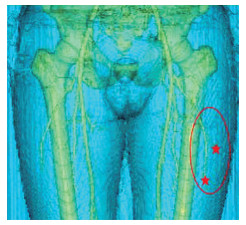
Clinical application of CTA combined with CDU-assisted anterolateral thigh flap in repairing and reconstructing defects after tongue cancer operation
-
摘要:
目的 舌癌术后组织缺损严重影响患者生活质量,要求在提高患者生存率的同时改善生活质量。笔者采用CT血管造影(CTA)、彩色多普勒超声(CDU)及二者联合的方式辅助设计股前外侧穿支皮瓣进行舌重建,寻求最少损伤获取舌缺损的最优重建。 方法 选取蚌埠医学院第一附属医院口腔科从2017年7月—2020年9月40例舌癌拟行手术治疗舌体缺损的患者,采用随机数字表法将患者随机分为3组,A组20例患者术前应用CTA联合CDU评估旋股外动脉的穿支走行等情况,B组10例患者术前应用CTA及C组10例患者应用CDU进行评估,术中同期行皮瓣修复术同时对3组术中穿支点符合率、穿支类型符合率均采用Fisher精确检验进行分析,手术时间、术后住院天数采用方差分析,P < 0.05为差异有统计学意义。 结果 37例患者皮瓣均存活,A组1例皮瓣部分坏死,肌瓣存活;B组及C组各有1例患者出现静脉危象抢救后仍坏死。A组与C组在穿支类型符合率方面差异存在统计学意义(P=0.001)且A组在皮瓣制备时间方面显著优于B、C 2组(P=0.004),3组在穿支点符合率(P=0.244)及术后住院天数(P=0.845)方面差异无统计学意义。术后供、受区切口一期愈合;患者舌部形态及功能等恢复均达到满意效果,供受区均未见明显并发症。 结论 CTA联合CDU对股前外侧皮瓣修复舌癌术后缺损具有较好的指导意义。 Abstract:Objective The postoperative tissue defect of tongue cancer seriously affects the quality of life of patients, which requires improvement of their survival rate and quality of life. CT angiography (CTA), colour Doppler ultrasound (CDU) and their combination were used to assist in designing anterolateral thigh perforator flap for tongue reconstruction so as to seek the best reconstruction of tongue defect with the least damage. Methods From July 2017 to September 2020, 40 patients with tongue cancer undergoing surgery for tongue defect were selected from the Department of Stomatology of the First Affiliated Hospital of Bengbu Medical College. The patients were randomly divided into three groups by random number table. Twenty patients in group A were evaluated by CTA combined with CDU before operation, 10 patients in group B were evaluated by CTA before operation and 10 patients in group C were evaluated by CDU before operation. Skin flap repair was performed at the same time during the operation. Fisher accurate probability method was used to analyse the coincidence rate of perforating fulcrum and perforating branch type in the three groups. Variance analysis was used to analyse the operation time and postoperative hospital stay, with P < 0.05 as the statistical significance. Results All the flaps of 37 patients survived. One case in group A was partially necrotic, but the muscle flap survived. One patient in group B and group C still died of venous crisis after rescue. A significant difference was observed between groups A and C in the coincidence rate of perforating branch types (P=0.001). The time of skin flap preparation in group A was significantly better than those in groups B and C (P=0.004), but no significant difference was noted among the three groups in the coincidence rate of perforating branch points (P=0.244) and postoperative hospitalization days (P=0.845). Regarding primary healing of donor and recipient incision after operation, satisfactory results were achieved in tongue shape and function recovery, and no obvious complications were found in donor and recipient areas. Conclusion CTA combined with CDU has a good guiding significance in repairing postoperative defects of tongue cancer with anterolateral thigh flap. -
表 1 3组舌癌患者一般资料比较[例(%)]
组别 例数 年龄(x ±s,岁) 性别 临床分期 男性 女性 Ⅱ Ⅲ Ⅳ A组 20 55.05±11.22 14(70.0) 6(30.0) 4(20.0) 8(40.0) 8(40.0) B组 10 55.70±14.15 6(60.0) 4(40.0) 4(40.0) 3(30.0) 3(30.0) C组 10 60.10±8.39 7(70.0) 3(30.0) 3(30.0) 4(40.0) 3(30.0) 统计量 0.682a 0.342b 1.027b P值 0.512 0.843 0.599 注:a为F值,b为χ2值。 表 2 3组舌癌患者手术情况比较
组别 例数 穿支点符合率[例(%)] 穿支类型符合率[例(%)] 皮瓣制备时间(x ±s,min) 术后住院天数(x ±s,d) A组 20 20(100.0) 20(100.0) 39.75±6.29 9.25±2.04 B组 10 9(90.0) 9(90.0) 46.20±4.29c 9.70±2.21 C组 10 9(90.0) 5(50.0)b 48.10±8.66c 9.50±1.90 统计量 6.564d 0.169d P值 0.244a 0.001a 0.004 0.845 注:a为采用Fisher精确检验;与A组比较,bP < 0.017(采用卡方分割,检验水准α校正=0.017),cP < 0.05;d为F值。 -
[1] ROBA T, CHEN Y Y, XU X H, et al. Short-term quality of life, functional status, and their predictors in tongue cancer patients after anterolateral thigh free flap reconstruction: a single-center, prospective, comparative study[J]. Cancer Manag Res, 2020, 12: 11663-11673. doi: 10.2147/CMAR.S268912 [2] 潘朝斌. 舌鳞癌的临床综合序列治疗研究进展[J]. 口腔疾病防治, 2018, 26(5): 273-280. https://www.cnki.com.cn/Article/CJFDTOTAL-GDYB201805001.htm [3] 常树森, 金文虎, 魏在荣, 等. 股前外侧皮瓣术前设计优化及临床应用[J]. 中华显微外科杂志, 2017, 40(2): 118-122. doi: 10.3760/cma.j.issn.1001-2036.2017.02.004 [4] 芮永军. 股前外侧皮瓣在中国的研究进展[J]. 中华显微外科杂志, 2020, 43(4): 313-325. doi: 10.3760/cma.j.cn441206-20200628-00277 [5] 莫勇军, 杨克勤, 谭海涛, 等. CTA联合彩色多普勒超声检测在股前外侧穿支皮瓣中的应用[J]. 中华显微外科杂志, 2018, 41(1): 68-72. doi: 10.3760/cma.j.issn.1001-2036.2018.01.017 [6] 郭宇, 魏在荣, 曾可为, 等. 高频彩色多普勒超声联合宽景成像在股前外侧穿支皮瓣术前导航中的应用[J]. 中国修复重建外科杂志, 2019, 33(2): 190-194. https://www.cnki.com.cn/Article/CJFDTOTAL-ZXCW201902015.htm [7] XIAO W, LI K, NG S K H, et al. A prospective comparative study of color doppler ultrasound and infrared thermography in the detection of perforators for anterolateral thigh flaps[J]. Ann Plast Surg, 2020, 84(5S): S190-S195. doi: 10.1097/SAP.0000000000002369 [8] 明华伟, 何芸, 张兴安, 等. CTA辅助游离股前外侧肌皮瓣修复舌癌术后缺损的应用研究[J]. 中国美容医学, 2019, 28(2): 23-26. https://www.cnki.com.cn/Article/CJFDTOTAL-MRYX201902011.htm [9] XIE R G. Medial versus lateral approach to harvesting of anterolateral thigh flap[J]. J Int Med Res, 2018, 46(11): 4569-4577. doi: 10.1177/0300060518786912 [10] 梁再卿, 吴宁. 股前外侧穿支皮瓣的临床应用研究进展[J]. 中华骨与关节外科杂志, 2019, 12(12): 1020-1024. doi: 10.3969/j.issn.2095-9958.2019.12.16 [11] STEVE A K, WhITE C P, ALKHAWAJI A, et al. Computed tomographic angiography used for localization of the cutaneous perforators and selection of anterolateral thigh flap"bail-out"branches[J]. Ann Plast Surg, 2018, 81(1): 87-95. doi: 10.1097/SAP.0000000000001433 [12] LU D, CHAN P, FERRIS S, et al. Anatomic symmetry of anterolateral thigh flap perforators: a computed tomography angiographic study[J]. ANZ J Surg, 2019, 89(5): 584-588. doi: 10.1111/ans.15005 [13] 邵侠, 屠呈威, 赵磊, 等. 股前外侧嵌合皮瓣与串联皮瓣修复口腔颌面部肿瘤根治术后缺损的效果比较[J]. 中华全科医学, 2018, 16(8): 1244-1246. https://www.cnki.com.cn/Article/CJFDTOTAL-SYQY201808006.htm [14] CARNEY M J, SAMRA F, MOMENI A, et al. Anastomotic technique and preoperative imaging in microsurgical lower-extremity reconstruction: a single-surgeon experience[J]. Ann Plast Surg, 2020, 84(4): 425-430. doi: 10.1097/SAP.0000000000002227 [15] 唐修俊, 王达利, 魏在荣, 等. 旋股外侧动脉降支皮瓣的个体化设计与供瓣区的生态保护[J]. 中华整形外科杂志, 2018, 34(7): 509-514. doi: 10.3760/cma.j.issn.1009-4598.2018.07.004 [16] GONZALEZ MARTINE J, TPRRES PEREA A, GIJON VEGA M, et al. Preoperative vascular planning of free flaps: comparative study of computed tomographic angiography, color doppler ultrasonography, and hand-held doppler[J]. Plast Reconstr Surg, 2020, 46(2): 227-237. http://journals.lww.com/plasreconsurg/Fulltext/2020/08000/Preoperative_Vascular_Planning_of_Free_Flaps_.3.aspx?Ppt=Article|plasreconsurg:2020:08000:00003|10.1097/prs.0000000000006966| [17] DE VIRGILIO A, IOCCA O, DI MAIO P, et al. Head and neck soft tissue reconstruction with anterolateral thigh flaps with various components: Development of an algorithm for flap selection in different clinical scenarios[J]. Microsurgery, 2019, 39(7): 590-597. doi: 10.1002/micr.30495 [18] LI K, LIN W, LI J W, et al. Reconstruction of tongue using anterolateral thigh free flap after radical surgery of tongue carcinoma[J]. Asian J Surg, 2020, 43(7): 775-776 doi: 10.1016/j.asjsur.2020.02.015 [19] 崔轶, 李国栋, 杨曦, 等. CTA联合手持彩色多普勒超声设计小腿穿支螺旋桨皮瓣的临床应用[J]. 中华显微外科杂志, 2019, 42(3): 232-236. doi: 10.3760/cma.j.issn.1001-2036.2019.03.006 [20] 李萌, 黄辉, 丁旭, 等. 术前彩色多普勒超声评估游离皮瓣供受区血管在头颈部修复重建中的初步应用研究[J]. 口腔医学, 2020, 40(5): 416-420. https://www.cnki.com.cn/Article/CJFDTOTAL-KQYX202005006.htm [21] 康永强, 吴永伟, 马运宏, 等. 穿支定位技术辅助股前外侧分叶穿支皮瓣移植修复前臂及手部大面积软组织缺损[J]. 中华创伤杂志, 2018, 34(10): 886-891. doi: 10.3760/cma.j.issn.1001-8050.2018.10.006 [22] 邹永通, 刘晓军, 练祝平, 等. 股前外侧穿支皮瓣移植在四肢严重创伤后皮肤缺损修复中的应用[J]. 海南医学, 2017, 28(11): 1854-1856. doi: 10.3969/j.issn.1003-6350.2017.11.046 -


 下载:
下载:


