

The relationship between the expression of HYAL1 and the progression and prognosis of head and neck squamous cell carcinoma
-
摘要:
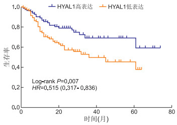
目的 本文旨在检查透明质酸酶1(HYAL1)在头颈鳞状细胞癌(HNSCC)中的表达水平及其与头颈鳞状细胞癌(HNSCC)患者的生存关系。 方法 收集2008—2015年浙江省人民医院行根治性切除的HNSCC患者资料构建组织芯片,通过免疫组织化学检测了258例患者切除的肿瘤标本和附近组织中HYAL1的表达。 结果 HYAL1在HNSCC组织中的表达明显低于正常邻近组织(P < 0.001)。HYAL1的低表达与肿瘤大小(P=0.011)、淋巴结转移(P=0.025)和TMN分期(P=0.038)显著相关。此外,与高HYAL1表达者相比,低HYAL1表达者与短生存期显著相关(P=0.007)。HYAL1低表达患者的总生存期中位数为36个月,而HYAL1高表达患者的总生存期仍未达到中位数(超过74个月)。在本研究有限的病例中,未发现高表达或低表达HYAL1的患者在年龄、性别、病变部位、HPV感染、吸烟、饮酒、组织学分期有任何差异。 结论 HYAL1低表达与HNSCC患者的预后不良有关。HYAL1可能是临床上有价值的预后指标,并且是HNSCC治疗的潜在靶标。 Abstract:Objective This article aims to study the expression level of hyaluronidase 1 (HYAL1) in head and neck squamous cell carcinoma (HNSCC) and its relationship with the survival of HNSCC patients. Methods The data of HNSCC patients who underwent radical resection in Zhejiang Provincial People's Hospital from 2008 to 2015 were collected to construct tissue chips, and the expression of HYAL1 in tumor specimens and nearby tissues from 258 patients was detected by immunohistochemistry. Results The expression of HYAL1 in HNSCC tissues was significantly lower than that of normal adjacent tissues (P < 0.001). The low expression of HYAL1 was significantly correlated with tumor size (P=0.011), lymph node metastasis (P=0.025) and TNMs staging system (P=0.038). In addition, compared with high HYAL1 expression, low HYAL1 expression was significantly associated with short survival time (P=0.007). The median overall survival of patients with low HYAL1 expression is 36 months, while the median overall survival of patients with high HYAL1 expression has not yet reached (more than 74 months). It is worth noting that in our limited cases, this study did not find any difference in age, gender, lesion site, HPV infection, smoking, drinking, and histological staging between the patients with high low expression of HYAL1. Conclusion The low expression of HYAL1 is related to the poor prognosis of these HNSCC patients. HYAL1 may be a clinically valuable prognostic indicator and a potential target for HNSCC treatment. -
Key words:
- Head and neck squamous cell carcinoma /
- Hyaluronidase 1 /
- Biomarker
-
表 1 HYAL1在头颈鳞状细胞癌中过表达(例)
组织类型 总计 HYAL1评分 χ2值 P值 高 低 头颈鳞状细胞癌 258 156 102 22.574 < 0.001 癌旁组织 132 111 21  下载: 导出CSV
表 2 HYAL1的表达与头颈鳞状细胞癌临床特征之间的关系(例)
项目 例数 HYAL1表达水平 χ2值 P值 高 低 性别 0.492 0.483 男性 203 125 78 女性 55 31 24 年龄(岁) 0.001 0.978 < 60 66 40 26 ≥60 192 116 76 肿瘤大小(cm) 9.531 0.002 ≤5 173 116 57 > 5 85 40 45 病变部位 5.847 0.664 牙龈 7 3 4 舌根 23 11 12 颊黏膜 36 16 20 口底 11 4 7 硬腭 14 7 7 咽部 8 4 3 唇 28 12 16 舌 36 10 26 喉 14 8 6 HPV感染 2.393 0.122 阳性 116 69 47 阴性 71 34 37 淋巴结转移 6.757 0.009 否 212 136 76 是 46 20 26 吸烟 0.648 0.421 是 153 88 65 否 64 33 31 TMN分期 8.439 0.004 Ⅰ~Ⅱ 201 131 70 Ⅲ~Ⅳ 57 25 32 组织学分期 4.206 0.122 高 53 21 32 中 132 72 60 低 25 15 10 饮酒 1.622 0.203 否 132 76 56 是 94 62 32
下载: 导出CSV
表 3 HNSCC患者临床病理参数和HYAL1表达的单因素Cox回归生存分析
项目 B SE Wald χ2 P值 HR(95% CI) HYAL1表达 1.166 0.681 7.472 0.009 0.574(0.329~0.902) 肿瘤大小 0.691 0.247 7.831 0.005 1.995(1.230~3.236) TMN分期 0.609 0.269 5.143 0.023 1.839(1.086~3.115) 组织学分期 0.345 0.309 1.246 0.017 2.134(1.260~3.628) 淋巴结转移 1.087 0.762 12.847 0.001 2.438(1.692~4.446) HPV感染 0.087 0.061 2.046 0.153 1.091(0.969~1.226)
下载: 导出CSV
表 4 HNSCC患者临床病理参数和HYAL1表达的多因素Cox回归生存分析
项目 B SE Wald χ2 P值 HR(95% CI) HYAL1表达 0.509 0.241 4.454 0.035 0.601(0.375~0.964) 肿瘤大小 0.337 0.212 2.515 0.113 1.401(0.924~2.124) TMN分期 1.595 0.326 23.970 < 0.001 4.929(2.603~9.335) 组织学分期 0.830 0.280 8.794 0.003 2.294(1.325~3.972) 淋巴结转移 1.053 0.247 18.225 < 0.001 2.868(1.768~3.651) HPV感染 0.097 0.242 0.161 0.689 1.102(0.685~1.772)
下载: 导出CSV
-
[1] ZHOU M, WANG H, ZENG X, et al. Mortality, morbidity, and risk factors in China and its provinces, 1990-2017: a systematic analysis for the Global Burden of Disease Study 2017[J]. Lancet, 2019, 394(10204): 1145-1158. doi: 10.1016/S0140-6736(19)30427-1 [2] LIU S, WOODY N, WEI W, et al. Evaluating compliance with process-related quality metrics and survival in oral cavity squamous cell carcinoma: Multi-institutional oral cavity collaboration study[J]. Head Neck, 2021, 43(1): 60-69. doi: 10.1002/hed.26454 [3] GOU Y, SHI M, YANG A, et al. Platinum-based chemotherapy plus cetuximab first-line for Asian patients with recurrent and/or metastatic squamous cell carcinoma of the head and neck: Results of an open-label, single-arm, multicenter trial[J]. Head Neck, 2015, 37(8): 1081-1087. doi: 10.1002/hed.23707 [4] CHANG Y, CHU L, LIU Y, et al. Verification of saliva matrix metalloproteinase-1 as a strong diagnostic marker of oral cavity cancer[J]. Cancers (Basel), 2020, 12(8): 2273-2290. doi: 10.3390/cancers12082273 [5] WICKER C, TAKIAR V, SUGANYA R, et al. Evaluation of antioxidant network proteins as novel prognostic biomarkers for head and neck cancer patients[J]. Oral Oncol, 2020, 111: 104949. doi: 10.1016/j.oraloncology.2020.104949 [6] 张宁, 兰雪玲. 铁蛋白在头颈癌颈部淋巴结转移的预测作用[J]. 实用肿瘤学杂志, 2019, 33(5): 442-446. https://www.cnki.com.cn/Article/CJFDTOTAL-SYZL201905014.htm [7] SPINELLI F, VITALE D, SEVIC I, et al. Hyaluronan in the tumor microenvironment[J]. Adv Exp Med Biol, 2020, 1245: 67-83. doi: 10.1007/978-3-030-40146-7_3 [8] DUAN H, DONOVAN M, HERNANDEZ F, et al. Hyaluronic-acid-presenting self-assembled nanoparticles transform a hyaluronidase HYAL1 substrate into an efficient and selective inhibitor[J]. Angew Chem Int Ed Engl, 2020, 59(32): 13591-13596. doi: 10.1002/anie.202005212 [9] LENG D, HUANG X, YI J, et al. HYAL1 is downregulated in idiopathic pulmonary fibrosis and inhibits HFL-1 fibroblast proliferation when upregulated[J]. Biomed Res Int, 2020, 2020: 3659451. http://www.researchgate.net/publication/339866044_HYAL1_Is_Downregulated_in_Idiopathic_Pulmonary_Fibrosis_and_Inhibits_HFL-1_Fibroblast_Proliferation_When_Upregulated [10] WITZEL I, MARX A, MULLER V, et al. Role of HYAL1 expression in primary breast cancer in the formation of brain metastases[J]. Breast Cancer Res Treat, 2017, 162(3): 427-438. doi: 10.1007/s10549-017-4135-6 [11] JIN Z, ZHANG G, LIU Y, et al. The suppressive role of HYAL1 and HYAL2 in the metastasis of colorectal cancer[J]. J Gastroenterol Hepatol, 2019, 34(10): 1766-1776. doi: 10.1111/jgh.14660 [12] 李志强, 姜书传, 马玲, 等. 膀胱移行癌中HYAL-1, CD44v6及MVD的临床意义[J]. 现代泌尿外科杂志, 2011, 16(5): 439-442. doi: 10.3969/j.issn.1009-8291.2011.05.018 [13] FRANZMANN E, SCHROEDER G, GOODWIN W, et al. Expression of tumor markers hyaluronic acid and hyaluronidase (HYAL1) in head and neck tumors[J]. Int J Cancer, 2003, 106(3): 438-445. doi: 10.1002/ijc.11252 [14] KAROUSOU E, MISRA S, GHATAK S, et al. Roles and targeting of the HAS/hyaluronan/CD44 molecular system in cancer[J]. Matrix Biol, 2017, 59: 3-22. doi: 10.1016/j.matbio.2016.10.001 [15] AGARWAL G, KRISHNAN K V, PRASAD S, et al. Biosynthesis of Hyaluronic acid polymer: Dissecting the role of sub structural elements of hyaluronan synthase[J]. Sci Rep, 2019, 9(1): 12510. doi: 10.1038/s41598-019-48878-8 [16] CHANMEE T, ONTONG P, ITANO N. Hyaluronan: A modulator of the tumor microenvironment[J]. Cancer Lett, 2016, 375(1): 20-30. doi: 10.1016/j.canlet.2016.02.031 [17] SUN S, WON H, HONG S, et al. Prognostic implications of stromal hyaluronic acid protein expression in resected oropharyngeal and oral cavity cancers[J]. Korean J Intern Med, 2020, 35(2): 408-420. doi: 10.3904/kjim.2018.203 [18] FROST G, MOHAPATRA G, WONG T, et al. HYAL1LUCA-1, a candidate tumor suppressor gene on chromosome 3p21.3, is inactivated in head and neck squamous cell carcinomas by aberrant splicing of pre-mRNA[J]. Oncogene, 2000, 19(7): 870-877. doi: 10.1038/sj.onc.1203317 [19] POSEY J, SOLOWAY M, EKICI S, et al. Evaluation of the prognostic potential of hyaluronic acid and hyaluronidase (HYAL1) for prostate cancer[J]. Cancer Res, 2003, 63(10): 2638-2644. http://cancerres.aacrjournals.org/cgi/reprint/63/10/2638 [20] CHENG X, WANG S, YANG H, et al. Negative regulation between the expression levels of receptor for hyaluronic acid-mediated motility and hyaluronan leads to cell migration in pancreatic cancer[J]. Oncol Lett, 2020, 20(5): 199-210. [21] MCATEE C, BOOTH C, ELOWSKY C, et al. Prostate tumor cell exosomes containing hyaluronidase Hyal1 stimulate prostate stromal cell motility by engagement of FAK-mediated integrin signaling[J]. Matrix Biol, 2019, 78: 165-179. http://www.sciencedirect.com/science?_ob=ShoppingCartURL&_method=add&_eid=1-s2.0-S0945053X1730464X&originContentFamily=serial&_origin=article&_ts=1526066327&md5=ba2d63a99b6b6460621fed0fcd6bb3c7 [22] NYKOPP T, PASONEN-SEPPANEN S, TAMMI M, et al. Decreased hyaluronidase 1 expression is associated with early disease recurrence in human endometrial cancer[J]. Gynecol Oncol, 2015, 137(1): 152-160. doi: 10.1016/j.ygyno.2015.01.525 [23] NAN H, YENER M, BUYRU N, et al. The investigation of hyaluronic acid and hyaluronidase-1 levels as tumour marker in larynx cancer[J]. Clin Otolaryngol, 2019, 44(6): 914-918. doi: 10.1111/coa.13390 [24] HUANG Q, BELZ G. Parallel worlds of the adaptive and innate immune cell networks[J]. Curr Opin Immunol, 2019, 58: 53-59. doi: 10.1016/j.coi.2019.04.008 [25] HAMADA M, YURA Y. Efficient delivery and replication of oncolytic virus for successful treatment of head and neck cancer[J]. Int J Mol Sci, 2020, 21(19): 7073-7094. doi: 10.3390/ijms21197073 -

 点击查看大图
图(1) / 表(4)
计量
- 文章访问数: 158
- HTML全文浏览量: 66
- PDF下载量: 1
- 被引次数: 0
