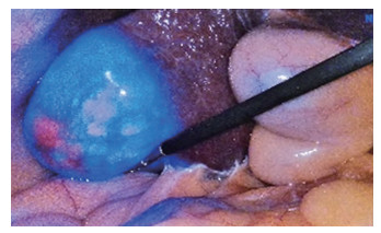
Application of ICG fluorescence imaging in laparoscopic hepatectomy for primary liver cancer
-
摘要:
目的 研究吲哚菁绿(indocyanine green,ICG)分子荧光显像技术在腹腔镜肝切除治疗原发性肝癌中的应用价值。 方法 回顾性分析2018年12月—2020年9月蚌埠医学院第一附属医院肝胆外科行ICG荧光腹腔镜肝切除术的56例原发性肝癌患者的临床及病理资料,统计分析手术方式、术前染色方法、术中荧光下肿瘤显影特点、新病灶的检测、手术后肿瘤病理结果等。 结果 所有患者均在ICG荧光腹腔镜下顺利完成肝切除手术,无中转开腹。有9例患者在荧光显像下出现新的可疑病灶,术中快速冰冻切片病理检查提示有4例证实为肝细胞性肝癌,1例为炎性改变,4例为硬化结节,9例患者中合并肝硬化患者8例。术中因肝硬化严重或交通支的存在导致10例患者染色失败,仅术前注射ICG的患者有8例染色失败,术中加行反染法的患者有2例染色失败。Child-Pugh分级为A级的患者在术前2 d或3 d给药可以在术中得到良好的显影效果,而B级的患者在术前5 d给药后荧光下肿瘤显影最佳。所有患者术后均无严重并发症发生,恢复良好。 结论 ICG荧光显像技术的发展为外科医生提供了一种简单有效的导航办法,可以准确定位肿瘤位置及切除边界,帮助发现浅表的微小病灶,但是具体如何界定肝硬化程度与注射时间仍需要讨论。 Abstract:Objective To study the application value of indocyanine green (ICG) molecular fluorescence imaging in laparoscopic hepatectomy for primary liver cancer. Methods The clinical and pathological data of 56 patients with primary liver cancer who underwent ICG fluorescence laparoscopic hepatectomy in the Department of Hepatobiliary Surgery of the First Affiliated Hospital of Bengbu Medical College from December 2018 to September 2020 were analysed retrospectively. The operative methods, the methods of preoperative staining, the characteristics of intraoperative fluorescence tumour development, the detection of new lesions and the pathological results of tumour after operation were statistically analysed. Results All patients successfully completed hepatectomy under ICG fluorescence laparoscopy without conversion to laparotomy. New suspicious lesions were found in 9 patients under fluorescence imaging. Pathological examination of intraoperative rapid frozen section showed 4 cases of hepatocellular carcinoma, 1 case of inflammatory changes and 4 cases of sclerotic nodules. Amongst the 9 patients, eight cases were complicated with liver cirrhosis. During the operation, staining failed in 10 patients due to severe liver cirrhosis or the presence of communicating branches. Staining failed in 8 patients who were injected with ICG before operation and 2 patients who received anti-staining during operation. The patients of Child-Pugh grade A could obtain a good development effect 2 or 3 days before operation, whereas those of grade B had the best tumour development under fluorescence 5 days before operation. All patients had no serious complications and recovered well. Conclusion The development of ICG fluorescence imaging technology provides surgeons with a simple and effective navigation method that can accurately locate the tumour and resection boundary and help to find superficial small lesions. However, how to define the degree of liver cirrhosis and injection time still need to be discussed. -
表 1 不同Child-Pugh分级术前注射时间情况(例)
级别 类别 2 3 4 5 6 A级 染色良好 18 20 染色欠佳 1 2 B级 染色良好 2 5 3 染色欠佳 3 0 2 注:Child-Pugh分级A级注射时间为2~3 d,B级注射时间为4~6 d。 表 2 术中新发病灶情况
病理类型 例数 肝硬化(例) 平均直径(cm) 平均深度(cm) HCC 4 3 0.90 0.36 硬化结节 4 4 0.63 0.28 炎性改变 1 1 0.60 0.10 -
[1] 郑荣寿, 孙可欣, 张思维, 等. 2015年中国恶性肿瘤流行情况分析[J]. 中华肿瘤杂志, 2019, 41(1): 19-28. doi: 10.3760/cma.j.issn.0253-3766.2019.01.005 [2] 王亦秋, 饶建华, 刘鹏, 等. 射频消融或微波消融分别联合肝动脉化疗栓塞治疗原发性肝癌的效果比较[J]. 中国临床研究杂志, 2017, 30(11): 1441-1445. https://www.cnki.com.cn/Article/CJFDTOTAL-ZGCK201711001.htm [3] 朱二畅, 鲁正, 徐建中, 等. 腹腔镜与开腹肝切除术治疗原发性肝细胞癌的临床疗效对比[J]. 中华全科医学, 2020, 18(11): 1845-1847,1973. https://www.cnki.com.cn/Article/CJFDTOTAL-SYQY202011015.htm [4] 中国研究型医院学会微创外科学专业委员会, 《腹腔镜外科杂志》编辑部. 吲哚菁绿荧光染色在腹腔镜肝切除术中应用的专家共识[J]. 腹腔镜外科杂志, 2019, 24(5): 388-394. https://www.cnki.com.cn/Article/CJFDTOTAL-FQJW201905023.htm [5] 中华人民共和国卫生和计划生育委员会医政医管局. 原发性肝癌诊疗规范(2017年版)[J]. 中华消化外科杂志, 2017, 16(7): 635-647. doi: 10.3760/cma.j.issn.1673-9752.2017.07.001 [6] NISHINO H, HATANO E, SEO S, et al. Real-time navigation for liver surgery using projection mapping with indocyanine green fluorescence: Development of the novel medical imaging projection system[J]. Ann Surg, 2018, 267(6): 1134-1140. doi: 10.1097/SLA.0000000000002172 [7] 张中林, 李晓勉, 李锟, 等. 吲哚菁绿荧光成像在腹腔镜肝脏外科手术中的应用[J]. 中华肝胆外科杂志, 2019, 25(2): 81-86. doi: 10.3760/cma.j.issn.1007-8118.2019.02.001 [8] 梁霄, 翟淑亭, 梁岳龙, 等. 荧光导航腹腔镜肝脏肿瘤切除吲哚菁绿术前给药时机: 单中心60例经验[J]. 中华肝胆外科杂志, 2019, 25(2): 90-93. doi: 10.3760/cma.j.issn.1007-8118.2019.02.003 [9] 董家鸿, 叶晟. 开启精准肝胆外科的新时代[J]. 中华普外科手术学杂志(电子版), 2016, 10(3): 181-184. doi: 10.3877/cma.j.issn.1674-3946.2016.03.001 [10] ZHANG Y, SHI R, HOU J C, et al. Liver tumor boundaries identified intraoperatively using real-time indocyanine green fluorescence imaging[J]. J Cancer Res Clin Oncol, 2017, 143(1): 51-58. doi: 10.1007/s00432-016-2267-4 [11] 方驰华, 梁洪玻, 迟崇巍, 等. 吲哚氰绿介导的近红外光技术在微小肝脏肿瘤识别, 切缘界定和精准手术导航的应用[J]. 中华外科杂志, 2016, 54(6): 444-450. doi: 10.3760/cma.j.issn.0529-5815.2016.06.011 [12] 王宏光. 吲哚菁绿肝段染色在腹腔镜肝癌切除中应用及意义[J]. 中国实用外科杂志, 2018, 38(4): 376-378. https://www.cnki.com.cn/Article/CJFDTOTAL-ZGWK201804010.htm [13] VAN MANEN L, HANDGRAAF H J M, DIANA M, et al. A practical guide for the use of indocyanine green and methylene blue in fluorescence-guided abdominal surgery[J]. J Surg Oncol, 2018, 118(2): 283-300. doi: 10.1002/jso.25105 [14] SUCHER R, BRUNOTTE M, SEEHOFER D. Indocyanine green fluorescence staining in liver surgery[J]. Chirurg, 2020, 91(6): 466-473. doi: 10.1007/s00104-020-01203-w [15] 张新龙, 刘杰. 荧光导航系统联合术中超声在精准腹腔镜肝肿瘤切除术中的应用[J]. 肝胆胰外科杂志, 2020, 32(6): 351-354. https://www.cnki.com.cn/Article/CJFDTOTAL-GDYW202006009.htm [16] 刘兵, 迟崇巍, 袁静, 等. 吲哚菁绿近红外荧光显像技术在肝细胞癌肝切除术中的应用价值[J]. 中华消化外科杂志, 2016, 15(5): 490-495. doi: 10.3760/cma.j.issn.1673-9752.2016.05.017 [17] TAKAHASHI H, ZAIDI N, BERBER E. An initial report on the intraoperative use of indocyanine green fluorescence imaging in the surgical management of the liver tumors[J]. J Surg Oncol, 2016, 114(5): 625-629. doi: 10.1002/jso.24363 [18] KAIBORI M, MATSUI K, ISHIZAKI M, et al. Intraoperative detection of superficial liver tumors by fluorescence imaging using indocyanine green and 5-aminolevulinic acid[J]. Anticancer Res, 2016, 36(4): 1841-1849. http://ar.iiarjournals.org/content/36/4/1841.full.pdf [19] KÖHN-GAONE J, GOGOI-TIWARI J, RAMM GRANT A, et al. The role of liver progenitor cells during liver regeneration, fibrogenesis, and carcinogenesis[J]. Am J Physiol Gastrointest Liver Physiol, 2016, 310(3): G143-G154. doi: 10.1152/ajpgi.00215.2015 [20] MIYATA A, ISHIZAWA T, TANI K, et al. Reappraisal of a dye-staining technique for anatomic hepatectomy by the concomitant use of indocyanine green fluorescence imaging[J]. J Am Coll Surg, 2015, 221(2): e27-36. doi: 10.1016/j.jamcollsurg.2015.05.005 [21] 王晓颖, 高强, 朱晓东, 等. 腹腔镜超声联合三维可视化技术引导门静脉穿刺吲哚菁绿荧光染色在精准解剖性肝段切除术中的应用[J]. 中华消化外科杂志, 2018, 17(5): 452-458. doi: 10.3760/cma.j.issn.1673-9752.2018.05.008 -


 下载:
下载:


