

Treatment of Judet Ⅳ type radial neck fracture in young children with Kirschner wire percutaneous leverage reduction and elastic intramedullary nail fixation under elbow arthrography
-
摘要:
目的 探讨肘关节造影下经皮克氏针撬拨复位后弹性髓内钉内固定方法在低龄儿童Judet Ⅳ型桡骨颈骨折治疗中的应用。 方法 选取安徽省儿童医院骨科2017年9月—2020年9月收治的骨折患儿29例,其中男11例,女18例,年龄3岁3个月~6岁11个月,均为外伤后1周内儿童Judet Ⅳ型桡骨颈骨折。均住院后在全麻下进行手术,选用碘海醇造影剂1 mL,混合生理盐水按照1∶ 1比例,肘后方间隙注入肘关节,C臂透视下可见原不显影的骨折断端软骨及骨骺,经皮克氏针撬拨骨折断端复位,自桡骨远端逆行穿入弹性髓内钉固定骨折断端,术后肘关节功能位石膏固定4周。 结果 本组29例患儿按Metaizeau的整复标准,其中优24例,良3例,可2例。本组29例患儿在门诊获得6~36个月的随访,参照Metaizeau疗效标准进行临床功能评定,优26例,良2例,可1例。本组患儿治疗优良率为96.5%,术后随访摄片未发现骨折再移位、桡骨小头坏死吸收等,所有患儿无骨折不愈合、骨化性肌炎、桡神经损伤、肘关节功能障碍等并发症。 结论 肘关节造影在低龄儿童Judet Ⅳ型桡骨颈骨折治疗中可清晰地显示原不显影的骨折断端软骨及骨骺。经皮克氏针撬拨复位创伤小,弹性髓内钉内固定骨折牢固,此方法尤其是对于治疗低龄儿童Judet Ⅳ型桡骨颈骨折效果好,远期关节功能恢复佳。 -
关键词:
- 肘关节造影 /
- 儿童 /
- Judet Ⅳ型桡骨颈骨折
Abstract:Objective To investigate the application of elastic intramedullary nail internal fixation after Kirschner wire (K-wire) percutaneous leverage reduction with elbow arthrography in the treatment of Judet Ⅳ type radial neck fracture in young children. Methods A total of 29 children (including 11 males and 18 females, aged 3 years and 3 months to 6 years and 11 months) in the Department of Orthopedics of Anhui Children's Hospital from September 2017 to September 2020 were selected. All children suffered Judet Ⅳ type radial neck fracture within one week after trauma. All patients underwent surgery under general anaesthesia after hospitalisation. The contrast agent was 1 mL iohexol, mixed with normal saline in a ratio of 1∶ 1 and injected into the elbow joint in the posterior space of the elbow. C-arm fluoroscopy showed the cartilage and epiphysis at the broken end of the fracture, which had not been developed before. The broken end of the fracture was reduced by K-wire pry, and the fracture was fixed by retrograde penetration of elastic intramedullary nail from the distal end of the radius. The functional position of the elbow was fixed in plaster for four weeks after surgery. Results According to Metaizeau's revision standard, 24 cases were excellent, 3 cases were good, and 2 cases were fair. The 29 children were followed up for 6-36 months in the outpatient department. According to Metaizeau's curative effect standard, 26 cases were excellent, 2 cases were good, and 1 case was fair. The excellent and good rate was 96.5%. Postoperative follow-up radiographs showed no fracture redisplacement and necrotic absorption of the radial head. No complications such as fracture nonunion, myositis ossificans, radial nerve injury and elbow joint dysfunction were observed in all cases. Conclusion Elbow arthrography can clearly show the cartilage and epiphysis at the broken end of the fracture without development in the treatment of the Judet Ⅳ type of radial neck fracture in young children. The trauma of reduction by K-wire prying is small, and the internal fixation by elastic intramedullary nail is firm. This method is especially effective for the treatment of Judet Ⅳ type radial neck fracture in young children, and the long-term joint function recovery is good. -
Key words:
- Elbow arthrography /
- Children /
- Judet Ⅳ type radial neck fracture
-

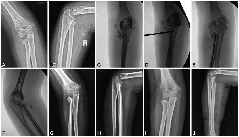
图 1 肘关节造影经皮克氏针撬拨复位弹性髓内钉内固定治疗Judet Ⅳ型桡骨颈骨折
注:患儿,男,6岁,Judet Ⅳ型桡骨颈骨折,A、B为骨折时肘关节正侧位片;C为术中肘关节造影透视片;D为克氏针撬拨复位后;E、F为弹性髓内钉内固定后;G、H为术后4周复查片;I、J为内固定取出4周后复查片。
-
[1] 张涛, 郭源, 吕学敏, 等. 2009—2016年28867例儿童肘部骨折的调查研究[J]. 骨科临床与研究杂志, 2018, 3(4): 218-224. https://www.cnki.com.cn/Article/CJFDTOTAL-GKLC201804008.htm [2] 刘玉昌, 王磊. 儿童桡骨颈骨折治疗技术及研究现状[J]. 实用骨科杂志, 2017, 23(6): 525-529. https://www.cnki.com.cn/Article/CJFDTOTAL-SGKZ201706014.htm [3] 王小波, 胡燕. 儿童肘关节损伤诊治失误原因分析[J]. 中国伤残医学, 2018, 26(8): 97-98. [4] ASHISH M, NARANG, ANUJA A, et al. Management of severely displaced radial neck fractures in children: A systematic review and meta-analysis of outcomes[J]. Indian J Orthop, 2020, 54(1): 60-68. doi: 10.1007/s43465-019-00032-2 [5] 吴声忠, 林廉洋, 周开亮, 等. 关节造影下微创治疗低龄儿童桡骨颈骨折21例[J]. 中国中医骨伤科杂志, 2020, 28(7): 75-77. https://www.cnki.com.cn/Article/CJFDTOTAL-ZGZG202007020.htm [6] SHABTAI L, ARKADER A. Percutaneous reduction of displaced radial neck fractures achieves better results compared with fractures treated by open reduction[J]. J Pediatr Orthop, 2016, 36: S63-S66. doi: 10.1097/BPO.0000000000000763 [7] 蔡英富, 崔顺平, 秦立亭. 双头加压螺钉及微型锁定接骨板在桡骨小头骨折内固定中的应用[J]. 海南医学, 2019, 30(19): 2507-2510. doi: 10.3969/j.issn.1003-6350.2019.19.018 [8] 杜香平, 余丽蓉, 熊志刚, 等. 克氏针经皮撬拨复位联合Métaizeau法治疗儿童Judet Ⅳ型桡骨颈骨折[J]. 中华手外科杂志, 2018, 34(1): 10-12. https://www.cnki.com.cn/Article/CJFDTOTAL-ZYZG201602009.htm [9] 林红明, 马一平, 胡成挺, 等. 改良Metaizeau法治疗JudetⅢ型或Ⅳ型儿童桡骨颈骨折[J]. 中国骨伤, 2018, 31(9): 790-793. doi: 10.3969/j.issn.1003-0034.2018.09.002 [10] 邓闽军, 闵继康, 徐旭纯, 等. 微创治疗儿童桡骨颈骨折[J]. 浙江临床医学, 2018, 20(4): 657-658. https://cdmd.cnki.com.cn/Article/CDMD-10285-1016221876.htm [11] 孙祥水, 侯华成, 汪飞, 等. 经皮血管钳捋拨复位弹性髓内针固定治疗儿童桡骨颈O'Brien Ⅱ、Ⅲ型骨折[J]. 中华小儿外科杂志, 2019, 40(2): 152-157. [12] 杨大兴, 单延具, 张琰, 等. 弹性髓内钉与克氏针固定治疗儿童桡骨颈骨折的临床对比[J]. 实用骨科杂志, 2019, 25(11): 961-964. https://www.cnki.com.cn/Article/CJFDTOTAL-SGKZ201911001.htm [13] 郭月超, 张玉舰, 王哲, 等. 弹性髓内针治疗儿童桡骨颈骨折的方案选择[J]. 河北医药, 2019, 41(9): 1391-1393, 1397. https://www.cnki.com.cn/Article/CJFDTOTAL-HBYZ201909028.htm [14] 朱佳, 夏先强, 赵诗铁, 等. 四肢骨折患者术后手术部位感染发生率及预测模型的构建[J]. 海南医学, 2021, 32(4): 456-459. https://www.cnki.com.cn/Article/CJFDTOTAL-HAIN202104013.htm [15] 王林, 陈梦婕, 王一臣, 等. 经皮撬拨辅助弹性髓内钉治疗小儿桡骨颈骨折的疗效研究[J]. 实用骨科杂志, 2019, 25(9): 830-832. https://www.cnki.com.cn/Article/CJFDTOTAL-SGKZ201909017.htm [16] 沈先涛, 陈小亮, 李雄涛, 等. B型超声引导经皮克氏针撬拨治疗儿童桡骨颈骨折[J]. 中华小儿外科杂志, 2015, 36(5): 363-367. [17] 姜海, 李敏, 吴永涛, 等. 术中肘关节造影辅助经皮撬拨复位克氏针内固定治疗儿童桡骨颈骨折[J]. 中国骨与关节杂志, 2017, 6(7): 517-521. https://www.cnki.com.cn/Article/CJFDTOTAL-GZGL201707012.htm [18] 姜海, 吴永涛, 汪兵, 等. 术中肘关节造影辅助治疗低龄儿童桡骨颈骨折[J]. 实用骨科杂志, 2019, 25(3): 197-200, 204. https://www.cnki.com.cn/Article/CJFDTOTAL-SGKZ201903002.htm [19] 袁毅, 金瑞, 姚杰, 等. 儿童闭合性Ⅲ型肱骨髁上骨折并桡神经损伤的疗效观察[J]. 中华全科医学, 2020, 18(2): 185-187, 276. https://www.cnki.com.cn/Article/CJFDTOTAL-SYQY202002006.htm [20] 胡成挺, 马一平. 弹性钉闭合复位内固定治疗儿童桡骨颈骨折[J]. 浙江临床医学, 2017, 19(3): 456-457. https://www.cnki.com.cn/Article/CJFDTOTAL-ZGZG202008014.htm [21] 易申德, 蔡军, 邹筠. 闭合复位弹性髓内钉固定和经皮克氏针撬拨复位固定治疗儿童桡骨颈骨折的疗效比较[J]. 实用临床医药杂志, 2021, 25(4): 77-80. https://www.cnki.com.cn/Article/CJFDTOTAL-XYZL202104020.htm [22] 陈星光, 刘珏, 王晓东, 等. 两种术式治疗儿童桡骨颈骨折疗效的Meta分析[J]. 中华小儿外科杂志, 2017, 38(5): 349-355. -

 下载:
下载:

 点击查看大图
图(2)
计量
- 文章访问数: 136
- HTML全文浏览量: 54
- PDF下载量: 1
- 被引次数: 0
