

MTP18 promotes oral squamous-cell carcinoma metastasis through induction of epithelial-mesenchymal transition
-
摘要:
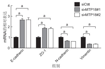
目的 探讨线粒体蛋白18(mitochondrial protein 18,MTP18)在口腔鳞癌转移中的调控作用。 方法 (1) 采用生物信息分析44例正常口腔黏膜与520例口腔鳞癌组织中MTP18的表达。(2)siRNA干涉MTP18表达后,用qRT-PCR与Western blotting实验检测干涉效果,用划痕与Transwell实验检测对细胞迁移与侵袭能力的影响。(3)siRNA下调MTP18表达后,用qRT-PCR实验分析对细胞上皮间质转化的影响。 结果 (1) 口腔鳞癌中MTP18表达显著上调(P<0.001),尤其在高级别与淋巴转移的肿瘤组织中。(2)siRNA显著下调了MTP18表达(P<0.001)。干涉MTP18可显著抑制口腔鳞癌SCC9细胞的迁移(siCtrl vs.siMTP18#1 vs. siMTP18#2为1.00±0.07 vs. 0.43±0.04 vs. 0.44±0.03,F=102.241, P<0.001)与侵袭(siCtrl vs. siMTP18#1 vs. siMTP18#2为1.00±0.07 vs. 0.43±0.04 vs. 0.44±0.03,F=97.017, P<0.001)。(3)干涉MTP18上调了上皮细胞标志分子的表达(P<0.001),而下调了间质细胞标志分子的表达(P<0.001)。 结论 MTP18通过诱导上皮间质转化促进口腔鳞癌细胞的迁移与侵袭。 Abstract:Objective To explore the biological functions of mitochondrial protein 18 (MTP18) in oral squamous-cell carcinoma metastasis. Methods (1) Bioinformatic analysis was conducted to evaluate the expression of MTP18 in 44 normal oral mucosa and 520 oral squamous-cell carcinoma tissues. (2) The silencing effect of MTP18 by siRNA was measured by qRT-PCR and Western blot analysis. In addition, the effects of MTP18 knockdown on the migration and invasion abilities of oral squamous-cell carcinoma were determined by wound-healing and Transwell invasion assays. (3) The effects of MTP18 knockdown on the expressions of markers in epithelial-mesenchymal transition (EMT) were determined by qRT-PCR analysis. Results (1) Bioinformatic analysis indicated that MTP18 expression was significantly increased in tumour tissues of oral squamous-cell carcinoma, especially in tumour tissues at high stages and with lymph node metastasis, compared with normal tissues (P < 0.001). (2) siRNA transfection successfully reduced the expression of MTP18 in oral squamous-cell carcinoma cells (P < 0.001). MTP18 knockdown significantly suppressed cell migration (siCtrl vs. siMTP18#1 vs. siMTP18#2: 1.00±0.07 vs. 0.43±0.04 vs. 0.44±0.03, F=102.241, P < 0.001) and invasion (siCtrl vs. siMTP18#1 vs. siMTP18#2: 1.00±0.07 vs. 0.43±0.04 vs. 0.44±0.03, F=97.017, P < 0.001). (3) MTP18 knockdown significantly increased the expressions of epithelial cell markers (P < 0.001), but suppressed the expressions of mesenchymal cell markers (P < 0.001). Conclusion MTP18 overexpression promotes oral squamous-cell carcinoma metastasis mainly through the induction of EMT. -
Key words:
- Mitochondrial protein 18 /
- Mitochondrial fission /
- Migration /
- Invasion /
- Oral squamous-cell carcinoma
-

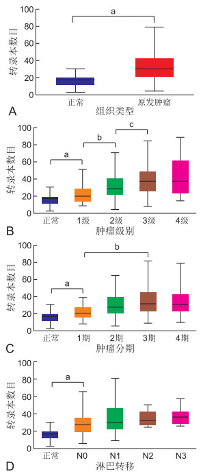
图 1 生物信息分析MTP18在口腔鳞癌中的表达变化
注:A为MTP18在癌组织与正常组织中表达;与正常组比较,aP < 0.001。B为MTP18在不同分级肿瘤中的表达;与正常组比较,aP < 0.001;与肿瘤分级为1级组比较,bP < 0.001;与肿瘤分级为2级组比较,cP < 0.001。C为MTP18在不同分期肿瘤中的表达;与正常组比较,aP < 0.001;与肿瘤分期为1期组比较,bP < 0.001。D为MTP18在转移性与非转移性肿瘤中的表达;与正常组比较,aP < 0.001。

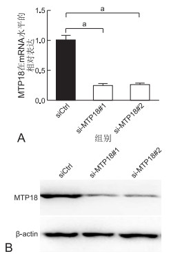
图 2 siRNA对口腔鳞癌SCC9细胞中MTP18表达的干涉效率分析
注:A为qRT-PCR;B为Western blotting。与siCtrl组比较,aP < 0.001。

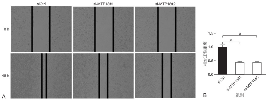
图 3 划痕实验分析下调MTP18对口腔鳞癌SCC9细胞迁移能力的影响
注:A为典型细胞划痕与迁移图片;B为各组细胞相对迁移距离的统计分析;与siCtrl组比较,aP < 0.001。

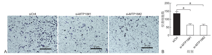
图 4 Transwell侵袭实验分析下调MTP18对口腔鳞癌SCC9细胞侵袭能力的影响
注:A为典型细胞侵袭图片;B为各组发生侵袭细胞数的统计分析;与siCtrl组比较,aP < 0.001。
-
[1] KOCH R E, JOSEFSON C C, HILL G E. Mitochondrial function, ornamentation, and immunocompetence[J]. Biol Rev Camb Philos Soc, 2017, 92(3): 1459-1474. doi: 10.1111/brv.12291 [2] STEREA A M, EL HIANI Y. The role of mitochondrial calcium signaling in the pathophysiology of cancer cells[J]. Adv Exp Med Biol, 2020, 1131(2): 747-770. doi: 10.1007/978-3-030-12457-1_30 [3] IDELCHIK M, BEGLEY U, BEGLEY T J, et al. Mitochondrial ROS control of cancer[J]. Semin Cancer Biol, 2017, 47(1): 57-66. [4] LOPEZ-LLUCH G, HERNANDEZ-CAMACHO J D, FERNANDEZ-AYALA D J M, et al. Mitochondrial dysfunction in metabolism and ageing: Shared mechanisms and outcomes?[J]. Biogerontology, 2018, 19(6): 461-480. doi: 10.1007/s10522-018-9768-2 [5] TILOKANI L, NAGASHIMA S, PAUPE V, et al. Mitochondrial dynamics: Overview of molecular mechanisms[J]. Essays Biochem, 2018, 62(3): 341-360. doi: 10.1042/EBC20170104 [6] NAN J, ZHU W, RAHMAN M S, et al. Molecular regulation of mitochondrial dynamics in cardiac disease[J]. Biochim Biophys Acta Mol Cell Res, 2017, 1864(7): 1260-1273. doi: 10.1016/j.bbamcr.2017.03.006 [7] FLIPPO K H, STRACK S. Mitochondrial dynamics in neuronal injury, development and plasticity[J]. J Cell Sci, 2017, 130(4): 671-681. http://www.onacademic.com/detail/journal_1000039814342010_b902.html [8] MISHRA P. Interfaces between mitochondrial dynamics and disease[J]. Cell Calcium, 2016, 60(3): 190-198. doi: 10.1016/j.ceca.2016.05.004 [9] SRINIVASAN S, GUHA M, KASHINA A, et al. Mitochondrial dysfunction and mitochondrial dynamics-The cancer connection[J]. Biochim Biophys Acta Bioenerg, 2017, 1858(8): 602-614. doi: 10.1016/j.bbabio.2017.01.004 [10] TROTTA A P, CHIPUK J E. Mitochondrial dynamics as regulators of cancer biology[J]. Cell Mol Life Sci, 2017, 74(11): 1999-2017. doi: 10.1007/s00018-016-2451-3 [11] KREYMERMAN A, BUICKIANS D N, NAHMOU M M, et al. MTP18 is a novel regulator of mitochondrial fission in CNS neuron development, axonal growth, and injury responses[J]. Sci Rep, 2019, 9(1): 10669. doi: 10.1038/s41598-019-46956-5 [12] ZHANG Y, LI H, CHANG H, et al. MTP18 overexpression contributes to tumor growth and metastasis and associates with poor survival in hepatocellular carcinoma[J]. Cell Death Dis, 2018, 9(10): 956. doi: 10.1038/s41419-018-0987-x [13] AUNG L H H, LI R, PRABHAKAR B S, et al. Mitochondrial protein 18 (MTP18) plays a pro-apoptotic role in chemotherapy-induced gastric cancer cell apoptosis[J]. Oncotarget, 2017, 8(34): 56582-56597. doi: 10.18632/oncotarget.17508 [14] CHANDRASHEKAR D S, BASHEL B, BALASUBRAMANYA S A H, et al. UALCAN: A portal for facilitating tumor subgroup gene expression and survival analyses[J]. Neoplasia, 2017, 19(8): 649-658. doi: 10.1016/j.neo.2017.05.002 [15] EL-HATTAB A W, SULEIMAN J, ALMANNAI M, et al. Mitochondrial dynamics: Biological roles, molecular machinery, and related diseases[J]. Mol Genet Metab, 2018, 125(4): 315-321. doi: 10.1016/j.ymgme.2018.10.003 [16] MA Y, WANG L, JIA R. The role of mitochondrial dynamics in human cancers[J]. Am J Cancer Res, 2020, 10(5): 1278-1293. http://www.ncbi.nlm.nih.gov/pubmed/32509379 [17] CHAN D C. Mitochondrial dynamics and its involvement in disease[J]. Annu Rev Pathol, 2020, 15(24): 235-259. [18] HUANG Q, ZHAN L, CAO H, et al. Increased mitochondrial fission promotes autophagy and hepatocellular carcinoma cell survival through the ROS-modulated coordinated regulation of the NFKB and TP53 pathways[J]. Autophagy, 2016, 12(6): 999-1014. doi: 10.1080/15548627.2016.1166318 [19] SUN X, CAO H, ZHAN L, et al. Mitochondrial fission promotes cell migration by Ca(2+) /CaMKII/ERK/FAK pathway in hepatocellular carcinoma[J]. Liver Int, 2018, 38(7): 1263-1272. doi: 10.1111/liv.13660 [20] ZHANG J, ZHANG Y, WU W, et al. Guanylate-binding protein 2 regulates Drp1-mediated mitochondrial fission to suppress breast cancer cell invasion[J]. Cell Death Dis, 2017, 8(10): e3151. doi: 10.1038/cddis.2017.559 [21] GHOSH A, CHATTERJEE K, CHOWDHURY A R, et al. Clinico-pathological significance of Drp1 dysregulation and its correlation to apoptosis in oral cancer patients[J]. Mitochondrion, 2020, 52(10): 115-124. http://www.sciencedirect.com/science/article/pii/S1567724919302296 [22] XIAO T, SUN J, XING Z, et al. MTFP1 overexpression promotes the growth of oral squamous cell carcinoma by inducing ROS production[J]. Cell Biol Int, 2020, 44(3): 821-829. doi: 10.1002/cbin.11278 [23] TANG H, PENG S, DONG Y, et al. MARCH5 overexpression contributes to tumor growth and metastasis and associates with poor survival in breast cancer[J]. Cancer Manag Res, 2019, 11(24): 201-215. http://www.onacademic.com/detail/journal_1000041685123899_a516.html -

 下载:
下载:
 点击查看大图
计量
- 文章访问数: 218
- HTML全文浏览量: 88
- PDF下载量: 3
- 被引次数: 0
