

Effect of preoperative total cholesterol and high-density lipoprotein-cholesterol levels on the prognosis of breast cancer
-
摘要:
目的 通过测定乳腺癌患者术前血清总胆固醇及高密度脂蛋白胆固醇水平,探讨术前总胆固醇及高密度脂蛋白胆固醇水平对乳腺癌患者预后的影响。 方法 回顾性收集2012年1月—2014年12月蚌埠医学院第一附属医院341例经病理确诊的乳腺癌患者清晨空腹多项血脂数据及临床病理资料,采用Kaplan-Meier分析法和Cox比例风险回归模型分析乳腺癌患者的总体生存期(OS)及无病生存期(DFS)。 结果 患者TC水平 < 6.2 mmol/L的平均OS为96.2个月,患者TC水平≥6.2 mmol/L的平均OS为95.4个月(P=0.556),患者TC水平 < 6.2 mmol/L的平均DFS为95.9个月,患者TC水平≥6.2 mmol/L的平均DFS为94.0个月(P=0.122)。在不同水平的TC组中,总体生存率及无病生存率差异均无统计学意义。患者HDL-C水平 < 1.04 mmol/L的平均OS为94.7个月,患者HDL-C水平≥1.04 mmol/L的平均OS为97.6个月(P=0.019),患者HDL-C水平 < 1.04 mmol/L的平均DFS为93.2个月,而患者HDL-C水平≥1.04 mmol/L的平均DFS为97.6个月(P=0.003)。在不同水平的HDL-C组中,总体生存期及无病生存期差异均有统计学意义。COX多因素分析发现,高水平HDL-C患者DFS更长(HR=3.916,95%CI:1.355~11.313, P=0.012)。 结论 术前HDL-C水平可能是预测乳腺癌患者DFS的独立因素。 Abstract:Objective To determine serum total cholesterol (TC) and high-density lipoprotein-cholesterol (HDL-C) of patients with breast cancer, explore the effect of preoperative blood lipid levels on the prognosis of patients with breast cancer. Methods Fasting plasma lipid in the morning and clinical pathological data of 341 patients with breast cancer confirmed by pathology from January 2012 to December 2014 in the First Affiliated Hospital of Bengbu Medical College were collected retrospectively. Kaplan-Meier analysis and Cox proportional hazard regression model were used to analyse overall survival (OS) and disease-free survival (DFS). Results The average OS of patients with TC < 6.2 mmol/L was 96.2 months, whereas that of patients with TC≥6.2 mmol/L was 95.4 months (P=0.556). The average DFS of patients with TC < 6.2 mmol/L was 95.9 months, and that of patients with TC≥6.2 mmol/L was 94.0 months (P=0.122). In different levels of TC groups, the OS rate and DFS rate were not statistically significant. The average OS of patients with HDL-C < 1.04 mmol/L was 94.7 months, whereas that of patients with HDL-C≥1.04 mmol/L was 97.6 months (P=0.019). The average DFS of patients with HDL-C < 1.04 mmol/L was 93.2 months, whereas the average DFS of patients with HDL-C level≥1.04 mmol/L was 94.7 months (P=0.003). In different levels of HDL-C groups, the OS time and DFS time were statistically significant. COX multivariate analysis showed that high HDL-C was beneficial to DFS (HR=3.916, 95% CI: 1.355-11.313, P=0.012). Conclusion The preoperative HDL-C level may be an independent factor predicting DFS in patients with breast cancer. -
Key words:
- Breast cancer /
- Blood lipid level /
- Prognosis
-

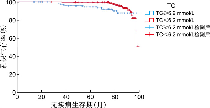
图 1 不同TC水平的总体生存期的K-M曲线
Figure 1. Overall survival curves of K-M at different levels of TC

图 2 不同TC水平的无疾病生存期的K-M曲线
Figure 2. Curves of disease-free survival with K-M at different levels of TC

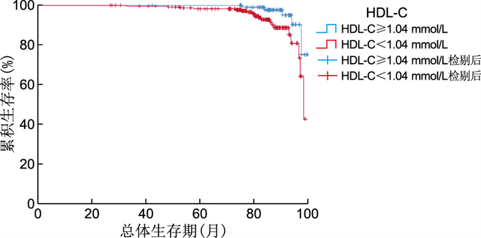
图 3 不同HDL-C水平的总体生存期的K-M曲线
Figure 3. Overall survival curves of K-M with different levels of HDL-C

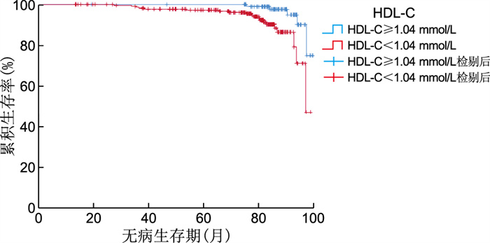
图 4 不同HDL-C水平的无疾病生存期的K-M曲线
Figure 4. Curves of disease-free survival with different levels of HDL-C and K-M
表 1 341例乳腺癌患者临床病理特征
Table 1. Clinicopathological features of 341 patients with breast cancer
项目 类别 例数 百分比(%) 年龄(岁) ≥45 202 59.2 < 45 139 40.8 月经状态 未绝经 138 40.5 绝经 203 59.5 肿瘤分期 T1 167 49.0 T2 153 44.9 T3 18 5.3 T4 3 0.9 淋巴结分期 N0 163 47.8 N1 88 25.8 N2 78 22.9 N3 12 3.5 分子分型 Luminal A型 61 17.9 Luminal B型 112 32.8 HER-2阳性型 86 25.2 TNBC型 82 24.0 TC(mmol/L) ≥6.2 110 32.3 < 6.2 231 67.7 TG(mmol/L) ≥2.3 90 26.4 < 2.3 251 73.6 HDL-C(mmol/L) ≥1.04 108 32.3 < 1.04 233 68.3 LDL-C(mmol/L) ≥3.4 113 33.1 < 3.4 228 66.9  下载: 导出CSV
表 2 341例乳腺癌患者不同TC水平与临床特征的关系(例)
Table 2. Relationship between different TC levels and clinical features in 341 breast cancer patients (cases)
项目 TC 统计量 P值 ≥6.2 mmol/L(n=110) < 6.2 mmol/L(n=231) 年龄 10.643a 0.001 ≥45岁 31 108 < 45岁 79 123 月经状态 13.355a 0.001 未绝经 60 78 绝经 50 153 肿瘤分期 7.935b 0.005 T1 45 122 T2 50 103 T3 13 5 T4 2 1 淋巴结分期 8.190b 0.004 N0 43 120 N1 28 60 N2 30 48 N3 9 3 分子分型 2.470a 0.481 Luminal A型 17 44 Luminal B型 34 78 HER-2阳性型 27 59 TNBC型 32 50 注:a为χ2值,b为H值。
下载: 导出CSV
表 3 341例乳腺癌患者不同HDL-C水平与临床特征的关系(例)
Table 3. Relationship between levels of different HDL-C and clinical features in 341 Patients with breast cancer (cases)
项目 HDL-C 统计量 P值 ≥1.04 mmol/L(n=108) < 1.04 mmol/L(n=233) 年龄 0.219a 0.640 ≥45岁 46 93 < 45岁 62 140 月经状态 2.530a 0.112 未绝经 37 101 绝经 71 132 肿瘤分期 14.800b < 0.001 T1 69 98 T2 36 117 T3 3 15 T4 0 3 淋巴结分期 0.550b 0.457 N0 52 111 N1 33 55 N2 21 57 N3 2 10 分子分型 3.385a 0.336 Luminal A型 24 37 Luminal B型 38 74 HER-2阳性型 23 63 TNBC型 23 59 注:a为χ2值,b为H值。
下载: 导出CSV
表 4 341例乳腺癌患者总体生存期、无疾病生存期的单因素Cox回归分析
Table 4. Univariate Cox regression analysis of overall survival and disease-free survival in 341 breast cancer patients
项目 OS单因素分析 DFS单因素分析 HR值 95% CI P值 HR值 95% CI P值 年龄 0.830 0.376~1.832 0.645 0.853 0.386~1.882 0.693 月经状态 0.649 0.293~1.436 0.286 0.635 0.287~1.402 0.261 分子分型 1.339 0.906~1.980 0.143 1.335 0.909~1.961 0.141 TC水平 0.780 0.340~2.387 0.557 0.525 0.229~1.204 0.128 TG水平 0.513 0.227~1.160 0.109 0.315 0.140~0.709 0.005 HDL-C水平 3.086 1.148~8.295 0.025 4.294 1.541~11.964 0.005 LDL-C水平 0.419 0.191~0.921 0.030 0.306 0.139~0.678 0.030
下载: 导出CSV
表 5 341例乳腺癌患者总体生存期、无疾病生存期多因素Cox比例分析
Table 5. Multivariate Cox ratio analysis of overall survival and disease-free survival in 341 breast cancer patients
项目 OS多因素分析 DFS多因素分析 HR值 95% CI P值 HR值 95% CI P值 TG水平 0.408 0.175~0.951 0.038 HDL-C水平 2.673 0.977~7.309 0.055 3.916 1.355~11.313 0.012 LDL-C水平 0.503 0.226~1.121 0.093 0.412 0.180~0.946 0.037
下载: 导出CSV
-
[1] SIEGEL R L, MILLER K D, JEMAL A. Cancer statistics, 2019[J]. CA Cancer J Clin, 2019, 69(1): 7-34. doi: 10.3322/caac.21551 [2] SUN Y S, ZHAO Z, YANG Z N, et al. Risk factors and preventions of breast cancer[J]. Int J Biol Med Sci, 2017, 13(11): 1387. doi: 10.7150/ijbs.21635 [3] KABAT G C, KIM M Y, CHLEBOWSKI R T, et al. Serum lipids and risk of obesity-related cancers in postmenopausal women[J]. Cancer Causes Control, 2018, 29(1): 13-24. doi: 10.1007/s10552-017-0991-y [4] NELSON E R. The significance of cholesterol and its metabolite, 27-hydroxycholesterol in breast cancer[J]. Mol Cell Endocrinol, 2018, 466: 73-80. doi: 10.1016/j.mce.2017.09.021 [5] 邓赢芳, 王淼舟. 血脂水平与乳腺癌关系的研究现状和进展[J]. 临床医药文献电子杂志, 2019, 6(5): 1. doi: 10.3877/j.issn.2095-8242.2019.05.001DENG Y F, WANG M Z. Research status and progress of the relationship between blood lipid level and breast cancer[J]. Journal of Clinical Medical Literature (ElectronicEdition), 2019, 6(5): 1. doi: 10.3877/j.issn.2095-8242.2019.05.001 [6] MAZZUFERI G, BACCHETTI T, ISLAM M O, et al. High density lipoproteins and oxidative stress in breast cancer[J]. Lipids Health Dis, 2021, 20(1): 1-13. doi: 10.1186/s12944-020-01429-x [7] TRYGVE L, MORTENSEN E S, HAWA N, et al. Impact of pre-diagnostic triglycerides and HDL-cholesterol on breast cancer recurrence and survival by breast cancer subtypes[J]. Bmc Cancer, 2018, 18(1): 1-11. doi: 10.1186/s12885-017-3892-2 [8] 中国抗癌协会乳腺癌专业委员会. 中国抗癌协会乳腺癌诊治指南与规范(2021年版)[J]. 中国癌症杂志, 2021, 31(10): 954-1040. https://www.cnki.com.cn/Article/CJFDTOTAL-ZGAZ202110015.htmBreast Cancer Committee of Chinese Anti-Cancer Association. Chinese Anti-Cancer Association Guidelines and Specifications for Diagnosis and Treatment of Breast Cancer (2021 edition)[J]. China Oncology, 2021, 31(10): 954-1040. https://www.cnki.com.cn/Article/CJFDTOTAL-ZGAZ202110015.htm [9] 中国成人血脂异常防治指南修订联合委员会. 中国成人血脂异常防治指南(2016年修订版)[J]. 中华健康管理学杂志, 2017, 11(1): 7-28. doi: 10.3760/j.issn.1674-0815.2017.01.003Joint Committee on the Revision of Guidelines for Prevention and Treatment of dyslipidemia in Adults in China. 2016 Chinese guideline for the management of dyslipidemia in adults[J]. Chinese Journal of Health Management, 2017, 11(1): 7-28. doi: 10.3760/j.issn.1674-0815.2017.01.003 [10] 潘婉婉, 董孟浩, 余发智, 等. 外周血炎症指标NLR、PLR、LMR预测乳腺癌新辅助化疗疗效的价值[J]. 中华全科医学, 2021, 19(9): 1442-1446. doi: 10.16766/j.cnki.issn.1674-4152.002081PAN W W, DONG M H, YU F Z, et al. Value of peripheral inflammatory markers NLR, PLR and LMR in predicting the efficacy of neoadjuvant chemotherapy for breast cancer[J]. Chinese general practice, 2021, 19(9): 1442-1446. doi: 10.16766/j.cnki.issn.1674-4152.002081 [11] NOWAK C, ǍRNLÖV J. A Mendelian randomization study of the effects of blood lipids on breast cancer risk[J]. Nat Commun, 2018, 9(1): 1-7. doi: 10.1038/s41467-017-02088-w [12] WANG Y Y, ATTANÉ C, MILHAS D, et al. Mammary adipocytes stimulate breast cancer invasion through metabolic remodeling of tumor cells[J]. JCI Insight, 2017, 2(4): e87489. DOI: 10.1172/jci.insight.87489. [13] 张艳利, 陈茂山, 杨光伦. 血清血脂和脂蛋白水平与乳腺癌发生及其临床病理学特征的相关性分析[J]. 中国普外基础与临床杂志, 2018, 25(2): 171-177. https://www.cnki.com.cn/Article/CJFDTOTAL-ZPWL201802009.htmZHANG Y L, CHEN M S, YANG G L. Association analysis of serum lipids and lipoprotein levels with the occurrence of breast cancer and clinicopathological characteristics of breast cancer patients[J]. Chinese Journal of Bases and Clinics in General Surgery, 2018, 25(2): 171-177. https://www.cnki.com.cn/Article/CJFDTOTAL-ZPWL201802009.htm [14] CHIMENTO A, CASABURI I, AVENA P, et al. Cholesterol and its metabolites in tumor growth: Therapeutic potential of statins in cancer treatment[J]. Front Endocrinol, 2019, 9: 807. doi: 10.3389/fendo.2018.00807 [15] MUNIR M T, PONCE C, POWELL C A, et al. The contribution of cholesterol and epigenetic changes to the pathophysiology of breast cancer[J]. J Steroid Biochem Mol Biol, 2018, 183: 1-9. doi: 10.1016/j.jsbmb.2018.05.001 [16] WU J, LEI X, PAN X, et al. Association between serum lipids and breast cancer risk in premenopausal women: Systematic review and meta-analysis[J]. J Int Med Res, 2021, 49(11). DOI: 10.1177/03000605211061033. [17] GUAITA-ESTERUELAS S, GUMA J, MASANA L, et al. The peritumoural adipose tissue microenvironment and cancer. The roles of fatty acid binding protein 4 and fatty acid binding protein 5[J]. Mol Cell Endocrinol, 2018, 462: 107-118. doi: 10.1016/j.mce.2017.02.002 [18] SHEIKHPOUR E, NOORBAKHSH P, FOROUGHI E, et al. A survey on the role of interleukin-10 in breast cancer: A narrative[J]. Rep Biochem Mol Biol, 2018, 7(1): 30-37. [19] 黄赞. 脂质代谢, 体质量指数与乳腺癌发生的关系[J]. 中国实用医药, 2019, 14(27): 3-5. https://www.cnki.com.cn/Article/CJFDTOTAL-ZSSA201927002.htmHUANG Z. Correlation of lipid metabolism, body mass index and breast cancer[J]. Practical medicine in China, 2019, 14(27): 3-5. https://www.cnki.com.cn/Article/CJFDTOTAL-ZSSA201927002.htm [20] OZMEN H K, ERDEMCI B, ASKIN S, et al. Carnitine and adiponectin levels in breast cancer after radiotherapy[J]. Open Med, 2017, 12: 189-194. doi: 10.1515/med-2017-0028 [21] 于子棋, 肖志, 邬玉辉, 等. 乳腺癌患者化疗前后血脂血糖及体质量指数的变化[J]. 中国普通外科杂志, 2017, 26(11): 1502-1505. doi: 10.3978/j.issn.1005-6947.2017.11.022YU Z Q, XIAO Z, WU Y H, et al. Changes of blood lipid, blood glucose and body mass index in breast cancer patients before and after chemotherapy[J]. Chinese Journal of General Surgery, 2017, 26(11): 1502-1505. doi: 10.3978/j.issn.1005-6947.2017.11.022 [22] ALOMAR S A, GǍMAN M A, PRABAHAR K, et al. The effect of tamoxifen on the lipid profile in women: A systematic review and meta-analysis of randomized controlled trials[J]. Exp Gerontol, 2022, 159: 111680. DOI: 10.1016/j.exger.2021.111680. [23] 李江涛. 研究阿那曲唑对乳腺癌术后化疗患者血脂、肝功能的影响[J]. 北方药学, 2018, 15(6): 29. https://www.cnki.com.cn/Article/CJFDTOTAL-BFYX201806020.htmLI J T. To study the effect of anastrozole on blood lipid and liver function in patients with breast cancer undergoing postoperative chemotherapy[J]. Journal of North Pharmacy, 2018, 15(6): 29. https://www.cnki.com.cn/Article/CJFDTOTAL-BFYX201806020.htm [24] JI Y L, ROUNDS T, CROCKER A, et al. The effect of atorvastatin on breast cancer biomarkers in high-risk women[J]. Cancer Prev Res, 2016, 9(5): 379-384. doi: 10.1158/1940-6207.CAPR-15-0300 -

 点击查看大图

计量
- 文章访问数: 251
- HTML全文浏览量: 175
- PDF下载量: 9
- 被引次数: 0
