

Application of human cartilage glycoprotein 39 and alpha-fetoprotein combined ultrasound in prenatal diagnosis of implanted placenta previa
-
摘要:
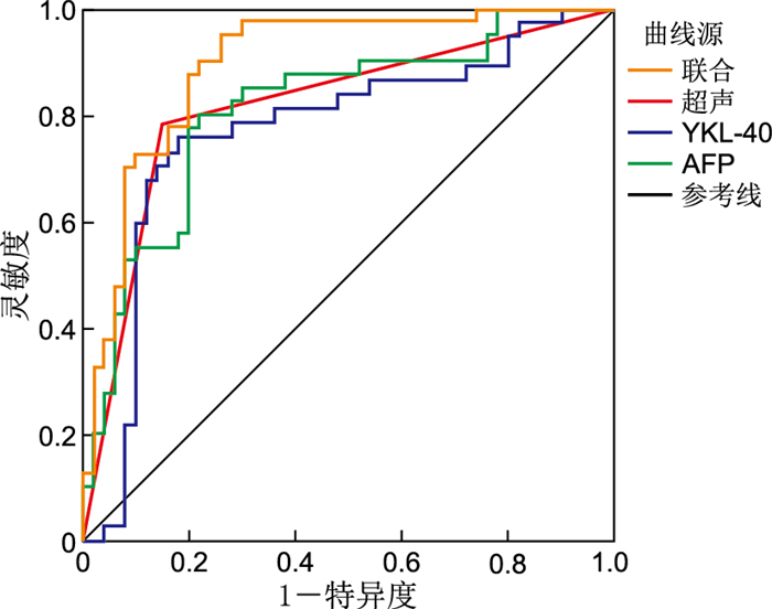
目的 探讨孕晚期人类软骨糖蛋白-39(YKL-40)、甲胎蛋白(AFP)联合超声对植入性前置胎盘的评估价值。 方法 收集2020年11月—2021年5月于蚌埠医学院第一附属医院分娩的前置胎盘孕妇90例为研究组,依据术中所见和(或)术后病理结果,分为植入组(穿透性4例,植入性17例,粘连性29例)50例和非植入组40例,并收集同期正常孕妇40例为对照组,入院均行YKL-40、AFP与超声检查。 结果 非植入组和对照组YKL-40、AFP水平低于植入组(均P<0.05),其中粘连性前置胎盘YKL-40低于植入性,植入性低于穿透性(均P<0.05),植入性和粘连性前置胎盘AFP低于穿透性(均P<0.05)。ROC曲线分析,血清YKL-40、AFP的最佳截断值分别为320.284 pg/mL、277.895 μg/mL,其曲线下的面积为0.780、0.812。联合诊断的AUC(0.895)、约登指数(0.675)、灵敏度(90.0%)、阴性预测值(85.3%)均高于单项检测。 结论 血清YKL-40、AFP水平联合超声有利于提高植入性前置胎盘的诊断效能。 -
关键词:
- 植入性前置胎盘 /
- 人类软骨糖蛋白-39 /
- 甲胎蛋白 /
- 超声
Abstract:Objective To evaluate the value of human cartilage glycoprotein-39 (YKL-40) and alpha fetoprotein (AFP) combined with ultrasound in placenta previa implantation. Methods A total of 90 pregnant women with placenta previa who delivered in the First Affiliated Hospital of Bengbu Medical College from November 2020 to May 2021 were enrolled as the research group. According to the intraoperative and/or postoperative pathological results, they were divided into the implantation group (4 cases of penetrating type, 17 cases of implantation type and 29 cases of adhesion type), 50 cases of non-implantation group and 40 cases of non-implantation group. In addition, 40 normal pregnant women in the same period were collected as the control group, and all received YKL-40, AFP and ultrasound examination. Results The levels of YKL-40 and AFP in the non-implantable group and control group were lower than those in the implantable group (all P < 0.05). The levels of YKL-40 in the implantable group were lower than those in the implantable group, and those in implantable group were lower than those in the penetrating group (all P < 0.05). AFP in the implantable group and implantable group were lower than those in the penetrating group (all P < 0.05). ROC curve analysis showed that the optimal cut-off values of serum YKL-40 and AFP were 320.284 pg/mL and 277.895 μg/mL, respectively, and the areas under the curve were 0.780 and 0.812, respectively. The AUC (0.895), Yuden index (0.675), sensitivity (90.0%) and negative predictive value (85.3%) of combined diagnosis were higher than those of single tests. Conclusion Serum YKL-40 and AFP levels combined with ultrasound can improve the diagnostic efficiency of placenta previa. -

图 1 AFP、YKL-40、超声及联合指标的ROC曲线
Figure 1. ROC curves of AFP, YKL-40, ultrasound and combined indexes
表 1 3组孕妇一般资料比较
Table 1. Comparison of general data among three groups of pregnant women
组别 例数 年龄(x±s,岁) 孕周(x±s,周) 孕次[M(P25, P75),次] 产次[M(P25, P75),次] 植入组 50 31.240±5.427 35.880±0.940 3.00(2.00, 5.00) 1.00(1.00, 1.00) 非植入组 40 31.225±4.633 36.250±0.981 3.00(2.00, 4.75) 1.00(0, 2.00) 对照组 40 31.050±5.277 36.225±1.025 3.00(2.00, 4.00) 1.00(0.25, 2.00) 统计量 0.018a 2.057a 0.251b 0.084b P值 0.983 0.132 0.882 0.976 注:a为F值,b为H值。  下载: 导出CSV
表 2 3组孕妇血清AFP、YKL-40比较(x±s)
Table 2. Comparison of serum AFP and YKL-40 in three groups(x±s)
组别 例数 AFP(μg/mL) YKL-40(pg/mL) 植入组 50 300.609±50.270 341.605±51.809 非植入组 40 253.032±36.440a 304.719±41.138a 对照组 40 255.790±11.943a 301.270±18.946a F值 23.069 13.842 P值 <0.001 <0.001 注:与植入组比较,aP < 0.05。
下载: 导出CSV
表 3 各类型植入组前置胎盘孕妇AFP、YKL-40水平比较(x±s)
Table 3. Comparison of AFP and YKL-40 levels in pregnant women with placenta previa in different implantation groups(x±s)
组别 例数 AFP(μg/mL) YKL-40(pg/mL) 穿透性 4 389.265±40.264 403.361±17.614 植入性 17 300.054±40.994a 358.001±49.294a 粘连性 29 288.706±44.929a 323.476±47.192ab F值 9.465 6.753 P值 <0.001 0.003 注:与穿透性比较,aP < 0.05;与植入性比较,bP < 0.05。
下载: 导出CSV
表 4 AFP、YKL-40、超声及联合指标的ROC结果
Table 4. ROC results of AFP, YKL-40, ultrasound and combined indexes
项目 AUC SE P值 95% CI 截点 AFP 0.812 0.047 <0.001 0.720~0.903 277.895 YKL-40 0.780 0.053 <0.001 0.675~0.884 320.284 超声 0.815 0.047 <0.001 0.722~0.908 联合 0.895 0.034 <0.001 0.828~0.962
下载: 导出CSV
-
[1] GIBBINS K J, EINERSON B D, VARNER M W, et al. Placenta previa and maternal hemorrhagic morbidity[J]. J Matern Fetal Neonatal Med, 2018, 31(4): 494-499. doi: 10.1080/14767058.2017.1289163 [2] JUNG E J, CHO H J, BYUN J M, et al. Placental pathologic changes and perinatal outcomes in placenta previa[J]. Placenta, 2018, 63: 15-20. doi: 10.1016/j.placenta.2017.12.016 [3] JAIN V, BOS H, BUJOLD E. Guideline No. 402: Diagnosis and management of placenta previa[J]. J Obstet Gynaecol Can, 2020, 42(7): 906-917. doi: 10.1016/j.jogc.2019.07.019 [4] JAUNIAUX E, KINGDOM J C, SILVER R M. A comparison of recent guidelines in the diagnosis and management of placenta accreta spectrum disorders[J]. Best Pract Res Clin Obstet Gynaecol, 2021, 72: 102-116. doi: 10.1016/j.bpobgyn.2020.06.007 [5] 郑言言, 张超学, 王玲, 等. 超声和磁共振成像在产前诊断胎盘植入中的应用价值比较[J]. 安徽医学, 2020, 41(10): 1164-1167. doi: 10.3969/j.issn.1000-0399.2020.10.013ZHENG Y Y, ZHANG X C, WANG L, et al. Comparison of ultrasound and magnetic resonance imaging in prenatal diagnosis of placenta accreta[J]. Anhui Medical Journal, 2020, 41(10): 1164-1167. doi: 10.3969/j.issn.1000-0399.2020.10.013 [6] JAUNIAUX E, COLLINS S, BURTON G J. Placenta accreta spectrum: Pathophysiology and evidence-based anatomy for prenatal ultrasound imaging[J]. Am J Obstet Gynecol, 2018, 218(1): 75-87. doi: 10.1016/j.ajog.2017.05.067 [7] 张茂春, 张红薇, 陈娇, 等. 产前超声征象评分联合肌酸激酶对前置胎盘合并胎盘植入的诊断价值分析[J]. 现代生物医学进展, 2018, 18(18): 3563-3567. https://www.cnki.com.cn/Article/CJFDTOTAL-SWCX201818037.htmZHANG M C, ZHANG H W, CHEN J, et al. Diagnostic value analysis of prenatal ultrasonographic signs score combined with creatine kinase for placenta previa combined with placenta implantation[J]. Progress in Modern Biomedicine, 2018, 18(18): 3563-3567. https://www.cnki.com.cn/Article/CJFDTOTAL-SWCX201818037.htm [8] PASQUO E D, GHI T, CALÌ G, et al. Intracervical lakes as sonographic marker of placenta accreta spectrum in patients with placenta previa and low-lying placenta[J]. Ultrasound Obset Gynecol, 2020, 55(4): 460-466. doi: 10.1002/uog.21866 [9] 刘艳梅, 刘红霞, 左春洁, 等. 超声图像特征在胎盘植入的产前诊断和产后子宫切除预测中的价值[J]. 陕西医学杂志, 2017, 46(4): 521-522. doi: 10.3969/j.issn.1000-7377.2017.04.051LIU Y M, LIU H X, ZUO C J, et al. Value of ultrasonographic features in prenatal diagnosis of placenta accreta and prediction of postpartum hysterectomy[J]. Shaanxi Medical Journal, 2017, 46(4): 521-522. doi: 10.3969/j.issn.1000-7377.2017.04.051 [10] 徐国华. 不同类型凶险性前置胎盘产妇的临床特点及剖宫产结局[J]. 中国妇幼保健, 2018, 33(9): 1986-1988. https://www.cnki.com.cn/Article/CJFDTOTAL-ZFYB201809022.htmXU G H. Clinical characteristics and cesarean section outcomes of different types of pernicious placenta previa[J]. Maternal and Child Health Care of China, 2018, 33(9): 1986-1988. https://www.cnki.com.cn/Article/CJFDTOTAL-ZFYB201809022.htm [11] 单华英, 费敬英, 查艺葆, 等. 前置胎盘孕妇产前超声在胎盘植入诊断中的价值[J]. 中华全科医学, 2017, 15(7): 1163-1165. doi: 10.16766/j.cnki.issn.1674-4152.2017.07.021SAN H Y, FEI J Y, ZA Y B, et al. Value of prenatal ultrasonography in diagnosis of placenta accreta in patients with placenta pracvia[J]. Chinese general practice, 2017, 15(7): 1163-1165. doi: 10.16766/j.cnki.issn.1674-4152.2017.07.021 [12] JAUNIAUX E, MOFFETT A, BURTUN G J. Placental implantation disorders[J]. Obstet Gynecol Clin North Am, 2020, 47(1): 117-132. doi: 10.1016/j.ogc.2019.10.002 [13] CHEN C, LIU X, CHEN D, et al. A risk model to predict severe postpartum hemorrhage in patients with placenta previa: A single-center retrospective study[J]. Ann Palliat Med, 2019, 8(5): 611-621. doi: 10.21037/apm.2019.09.04 [14] 张超, 王娜, 安丽, 等. 超声联合肌酸激酶及AFP检查凶险型前置胎盘合并胎盘植入的诊断和预后评估价值分析[J]. 河北医药, 2018, 40(13): 1940-1944. doi: 10.3969/j.issn.1002-7386.2018.13.004ZHANG C, WANG N, AN L, et al. The application value of ultrasound combined with detection of creatine kinase and AFP in diagnosis and prognosis evaluation of dangerous placenta previa complicated by placenta accreta[J]. Hebei Medicine, 2018, 40(13): 1940-1944. doi: 10.3969/j.issn.1002-7386.2018.13.004 [15] KONG C W, TO W W K. Risk factors for severe postpartum haemorrhage during caesarean section for placenta praevia[J]. J Obstet Gynaecol, 2020, 40(4): 479-484. doi: 10.1080/01443615.2019.1631769 [16] BERKLEY E M, ABUNHAMAD A. Imaging of placenta accreta spectrum[J]. Clin Obstet Gynecol, 2018, 61(4): 755-765. doi: 10.1097/GRF.0000000000000407 [17] RECKLIES A D, WHITE C, LING H. The chitinase 3-like protein human cartilage glycoprotein 39(HC-gp39)stimulates proliferation of human connective-tissue cells and activates both extracellular signal-regulated kinase-and protein kinase B-mediated signalling pathways[J]. Biochem J, 2002, 365(Pt1): 119-126. [18] DE CEUNINCK F, GAUFILLIER S, BONNAUD A, et al. YKL-40(cartilage gp-39) induces proliferative events in cultured chondrocytes and synoviocytes and increases glycosaminoglycan synthesis in chondrocyes[J]. Biochem Biophys Res Commun, 2021, 285(4): 926-931. [19] GÖZÜKARA Ï, ÖZGÜR T, DOLAPÇIOĜLU K, et al. YKL-40 expression in abnormal invasive placenta cases[J]. J Perinat Med, 2017, 45(5): 571-575. [20] 李泽丽, 陈练, 赵扬玉. 分子标志物在胎盘植入性疾病产前诊断中的应用价值[J]. 中国实用妇科与产科杂志, 2021, 37(11): 1105-1108. https://www.cnki.com.cn/Article/CJFDTOTAL-ZGSF202111007.htmLI Z L, CHEN L, ZHAO Y Y. Application value of molecular biomarkers in prenatal diagnosis of placenta accreta spectrum disorders[J]. Chinese Journal of Practical Gynecology and Obstetrics, 2021, 37(11): 1105-1108. https://www.cnki.com.cn/Article/CJFDTOTAL-ZGSF202111007.htm -

 点击查看大图
计量
- 文章访问数: 161
- HTML全文浏览量: 77
- PDF下载量: 0
- 被引次数: 0
