Inner ear malformation among 255 cases with severe to extremely severe sensorineural hearing loss in Xinjiang and relevant issues
-
摘要:
目的 探讨重度-极重度感音神经性耳聋患儿的内耳畸形的影像及手术特点,从而更好地为人工耳蜗植入提供临床指导。 方法 调取新疆维吾尔自治区人民医院2020年1月—2021年12月筛查并行人工耳蜗植入术的内耳畸形患儿的颞骨高分辨率CT资料,根据Sennaroglu的分类方法对其进行分类,依次为Michel畸形、耳蜗未发育、共同腔畸形、耳蜗发育不良(Ⅰ型、Ⅱ型、Ⅲ型)、不完全分隔Ⅰ型(IP-Ⅰ)、Ⅱ型(IP-Ⅱ)、Ⅲ型(IP-Ⅲ)、前庭及半规管畸形、前庭导水管扩大及内听道狭窄等,并记录人工耳蜗植入径路、电极类型及并发症,分别分析各类畸形的影像特点及手术注意事项。 结果 255例重度-极重度感音神经性耳聋患者中有66例(125耳)内耳畸形患者,其中,IP-Ⅱ型占内耳畸形的30.30%(20例/66例),单纯前庭导水管扩大占19.70%(13例/66例),其他类型比例较低。共55例(57耳)内耳畸形患儿接受了人工耳蜗植入手术,双侧植入2例。所有畸形病例均鼓阶开窗或圆窗植入电极。耳蜗发育不良(Ⅱ型、Ⅲ型)、IP-Ⅰ和IP-Ⅲ型选择短的直电极,IP-Ⅱ型、前庭导水管扩大、内听道扩大者选择标准电极。1例IP-Ⅱ患者术后CT提示植入前庭,二次手术植入耳蜗。“井喷”发生率为29.82% (17耳/57耳)。无脑脊液耳漏、皮瓣坏死等并发症。 结论 内耳畸形中IP-Ⅱ和大前庭导水管(LVA)两型占主导,内耳畸形患儿人工耳蜗植入术中“井喷”发生率高,高分辨率CT与MRI互补,可清晰、全面地显示内耳结构,为人工耳蜗植入手术的顺利实施提供保障。 Abstract:Objective To investigate the image and surgical characteristics of inner ear malformation in children with severe to extremely severe sensorineural hearing loss, so as to provide better clinical guidance for cochlear implantation. Methods From January 2020 to December 2021, children admitted to People's Hospital of Xinjiang Uygur Autonomous Region for screening and cochlear implantation were examined by high-resolution CT of temporal bone and the cases of inner ear malformation were screened out. According to Sennaroglu ' s classification method, they were classified as follows: Michel malformation, cochlea aplasia, common cavity, cochlear hypoplasia types Ⅰ, Ⅱ and Ⅲ, incomplete partition types Ⅰ-Ⅲ (IP-Ⅰ, IP-Ⅱ and IP-Ⅲ), vestibular and semicircular canal malformation, large vestibular aqueduct and internal auditory canal stenosis. Cochlear implantation approaches, electrode types and complications were recorded, and the image characteristics and surgical precautions of various malformations were analysed. Results Among 255 patients with severe to extremely severe sensorineural hearing loss, 66 cases (125 ears) had inner ear malformation, in which IP-Ⅱ accounted for 30.30% (20 cases/66 cases) of inner ear malformation cases, large vestibular aqueduct accounted for 19.70% (13 cases/66 cases) and other types were relatively low. A total of 55 children (57 ears) with inner ear malformation received cochlear implantation (CI), including two bilateral CI cases. All cases had been implanted with electrodes through a cochleotomy or round window. Short straight electrodes were implanted in patients with cochlear hypoplasia types Ⅱ and Ⅲ, IP-Ⅰ and IP-Ⅲ. Standard electrodes were implanted in patients with IP-Ⅱ, large vestibular aqueduct and enlarged internal auditory canal. Postoperative CT scans suggested one IP-Ⅱ case were implanted into vestibular cavity, so this case received another operation by cochlear implantation successfully. The incidence of cerebrospinal fluid "gusher" was 29.82% (17 ears/57 ears). There was no complications such as cerebrospinal fluid otorrhea and flap necrosis. Conclusion IP-Ⅱ and LVA are the predominant types of inner ear malformations. The incidence of cerebrospinal fluid "gusher" in CI is high in children with inner ear malformation. HRCT and MRI are complementary, which can clearly and comprehensively display the inner ear structure and provide guarantee for cochlear implementation successfully. -
Key words:
- Inner ear malformation /
- High-resolution CT /
- Sensorineural hearing loss
-
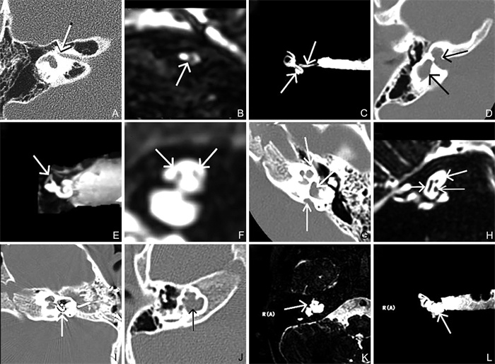
图 1 病例一至病例四影像学资料
注:病例一,A中白箭头示右侧内听道骨性狭窄;B中白箭头示右侧前庭下神经纤细,耳蜗神经未显示;C中上方白箭头示狭窄的内听道,下方箭头示耳蜗及前庭水成像结构正常;病例二,D示囊状耳蜗,无蜗轴,伴囊性前庭;E中左侧白箭头示面神经,右侧白箭头示前庭神经,耳蜗神经未显示;F示上半规管显示,前庭扩大;病例三,G示中间周和顶周融合呈囊状,顶部的蜗轴和分隔缺如,伴有前庭及前庭导水管的扩大;H中左侧白箭头示前庭神经,右上白箭头示面神经,右下白箭头示耳蜗神经;I示IP-Ⅱ型畸形,人工耳蜗植入时电极插入前庭腔;病例四,J中白箭头示耳蜗呈塔状,蜗轴缺如;K示耳蜗有分隔,无蜗轴;L示前庭扩大,上半规管、后半规管可见。
Figure 1. Imaging examination of case 1
表 1 255例重度-极重度感音神经性耳聋患者的一般情况
Table 1. General characteristics of 255 patients with severe sensorineural hearing loss
组别 例数 畸形数(耳) 年龄[M(P25, P75), 岁] CI数(耳) 井喷率(%) 畸形组 66 125 5(3, 6) 57 29.82(17/57) 正常组 189 0 5(3, 6) 190 2.65(5/190)a 合计 255 125 5(3, 6) 247 8.91(22/247) 注:2组井喷率比较,χ2=39.961,aP<0.001。 表 2 内耳畸形患儿CI植入情况
Table 2. Characteristics of cochlear implantation in patients with inner ear malformation
类型 耳数 CI植入耳数 井喷例数 并发症例数 CH 8 3 0 0 IP-Ⅰ 6 2 2 0 IP-Ⅱ 40 21 7 1 IP-Ⅲ 4 2 2 0 LVA 26 15 6 0 内听道畸形 9 4 0 0 前庭及半规管畸形 14 7 0 0 合计 107 54 17 1 -
[1] DAHLEN R T, HARNSBERGER H R, GRAY S D, et al. Overlapping thin-section fast spin-echo MR of the large vestibular aqueduct syndrome[J]. AJNR Am J Neuroradiol, 1997, 18(1): 67-75. [2] 刘军, 李长勤. 微小内耳畸形的感音神经性耳聋患儿与正常儿童内耳HRCT标准化测量的对比研究[J]. 医学影像学杂志, 2016, 26(11): 1962-1965. https://www.cnki.com.cn/Article/CJFDTOTAL-XYXZ201611005.htmLIU J, LI C Q. The comparative study of small inner ear malformation of sensorineural hearing loss children and norraul children of the comparison[J]. J Med Imaging, 2016, 26(11): 1962-1965. https://www.cnki.com.cn/Article/CJFDTOTAL-XYXZ201611005.htm [3] 鲁兆毅, 王宇, 潘滔. 听神经发育异常合并中耳内耳畸形及面神经畸形的人工耳蜗植入术1例[J]. 中华耳科学杂志, 2018, 16(6): 861-863. doi: 10.3969/j.issn.1672-2922.2018.06.022LU Z Y, WANG Y, PAN T. Cochlear implantation in a patient with auditory nerve hypoplasia, middle and inner ear malformation and facial nerve malformation: A case report[J]. Chinese Journal of Otology, 2018, 16(6): 861-863. doi: 10.3969/j.issn.1672-2922.2018.06.022 [4] 马辉, 韩萍, 梁波, 等. 多层螺旋CT对先天性内耳发育畸形的诊断价值[J]. 中华耳鼻咽喉头颈外科杂志, 2005, 40(4): 275-278. doi: 10.3760/j.issn:1673-0860.2005.04.009MA H, HAN P, LIANG B, et al. Diagnostic significance of multi-slice computed tomography imaging in congenital inner ear malformations[J]. Chin J Otorhinolaryngol Head Neck Surg, 2005, 40(4): 275-278. doi: 10.3760/j.issn:1673-0860.2005.04.009 [5] 鲜军舫, 王振常, 燕飞, 等. MRI快速自旋回波T2WI三维重建技术在内耳病变中的应用[J]. 中华放射学杂志, 1999, 33(7): 473-476. doi: 10.3760/j.issn:1005-1201.1999.07.009XIAN J F, WANG Z C, YAN F, et al. Application of MRI fast spin echo T2Wl 3D reconstruction in inner ear lesions[J]. Chinese Journal of Radiology, l999, 33(7): 473-476. doi: 10.3760/j.issn:1005-1201.1999.07.009 [6] 李建红, 王振常, 鲜军舫, 等. MRI在先天性内耳畸形儿童人工耳蜗植入术前的评估价值[J]. 磁共振成像, 2012, 3(6): 415-419. https://www.cnki.com.cn/Article/CJFDTOTAL-CGZC201206007.htmLI J H, WANG Z C, XIAN J F, et al. Value of MRI in evaluating the children with congenital inner ear malformations before cochlear implantation[J]. Chin J Magn Reson Imaging, 2012, 3(6): 415-419. https://www.cnki.com.cn/Article/CJFDTOTAL-CGZC201206007.htm [7] 丁贺宇, 赵鹏飞, 吕晗, 等. 先天性感音神经性耳聋面神经管迷路段与耳蜗间骨壁的HRCT研究[J]. 临床放射学杂志, 2019, 38(2): 229-232. https://www.cnki.com.cn/Article/CJFDTOTAL-LCFS201902010.htmDING H Y, ZHAO P F, LV H, et al. High-resolution CT study on the bony septum between labyrinth segment of facial nerve canal and cochlear in congenital sensorineural hearing loss[J]. Chin J Magn Reson Imaging, 2019, 38(2): 229-232. https://www.cnki.com.cn/Article/CJFDTOTAL-LCFS201902010.htm [8] 周宝, 林少莲, 林有辉, 等. 先天性内耳畸形的影像学及听力学分析[J]. 临床耳鼻咽喉头颈外科杂志, 2015, 29(22): 1950-1954. https://www.cnki.com.cn/Article/CJFDTOTAL-LCEH201522005.htmZHOU B, LIN S L, LING Y H, et al. Imaging and audiology analysis of the congenital inner ear malformations[J]. J Clin Otorhinolaryngol Head and Neck Surg, 2015, 29(22): 1950-1954. https://www.cnki.com.cn/Article/CJFDTOTAL-LCEH201522005.htm [9] KUMAR S, MAJHI B N, YADAV K K, et al. Radiological anatomy of inner ear malformation in hearing impaired children and it ' s correlation with hearing loss: A hospital based observational study[J]. Indian J Otolaryngol Head Neck Surg, 2018, 70(2): 278-283. doi: 10.1007/s12070-017-1238-7 [10] SENNAROGLU L. A new classification for cochleovestibular malformations[J]. Laryngoscope, 2002, 112(12): 2230-2241. doi: 10.1097/00005537-200212000-00019 [11] 姜泗长. 耳科学[M]. 上海: 上海科学技术出版社, 2002: 24-33.JIANG S C. Otology[M]. Shanghai: Shanghai Science and Technology Press, 2002: 24-33. [12] SNOW J B, WACKYM P A. Ballenger耳鼻咽喉头颈外科学[M]. 北京: 人民卫生出版社, 2012: 25-28.SNOW J B, WACKYM P A. Ballenger otorhinolaryngology head neck surgery[M]. Beijing: People ' s Medical Publishing House, 2012: 25-28. [13] LIM R, BRICHTA A M. Anatomical and physiological development of the human inner ear[J]. Hear Res, 2016, 338(8): 9-21. [14] 孙宝春. 内耳畸形影像学诊断及分类的研究进展[J]. 医学信息(中旬刊), 2011, 24(5): 1958-1959. https://www.cnki.com.cn/Article/CJFDTOTAL-YXSS201105328.htmSUN B C. Advances in imaging diagnosis and classification of inner ear malformations[J]. Medical Information, 2011, 24(5): 1958-1959. https://www.cnki.com.cn/Article/CJFDTOTAL-YXSS201105328.htm [15] JACKLER R K, CRUZ A D L. The large vestibular aqueduct syndrome[J]. Laryngoscope, 1989, 99(12): 1238-1242. [16] GOLDFELD M, GLASER B, NASSIR E, et al. CT of the ear in Pendred syndrome[J]. Radiology, 2005, 235(2): 537-540. [17] 耿国兴, 林彩娟, 黄小桃, 等. 42 708例新生儿耳聋基因筛查结果分析[J]. 中华全科医学, 2020, 18(10): 1688-1690. doi: 10.16766/j.cnki.issn.1674-4152.001594GENG G X, LING C J, HUANG X T, et al. An analysis of deafness genes screening in 42 708 newborns[J]. Chinese Journal of General Practice, 2020, 18(10): 1688-1690. doi: 10.16766/j.cnki.issn.1674-4152.001594 [18] 刘兰, 费静, 陶美慧, 等. 内耳发育畸形患者人工耳蜗植入术后的疗效分析[J]. 中国耳鼻咽喉颅底外科杂志, 2019, 25(2): 152-156. https://www.cnki.com.cn/Article/CJFDTOTAL-ZEBY201902013.htmLIU L, FEI J, TAO M H, et al. Efficacy of cochlear implantation in patients with inner ear malformations[J]. Chinese Journal of Otorhinolaryngology-skull Base Surgery, 2019, 25(2): 152-156. https://www.cnki.com.cn/Article/CJFDTOTAL-ZEBY201902013.htm [19] 陈艳丹, 陈柳, 华清泉. 内耳发育畸形的人工耳蜗植入术[J]. 中国医药导报, 2017, 14(12): 113-116. https://www.cnki.com.cn/Article/CJFDTOTAL-YYCY201712028.htmCHEN Y D, CHEN L, HUA Q Q. Cochlear implantation of the inner ear malformations[J]. China Medical Hearld, 2017, 14(12): 113-116. https://www.cnki.com.cn/Article/CJFDTOTAL-YYCY201712028.htm [20] MATHIS J M, SIMMONS D M, HE X, et al. Brain 4: A novel mammalian POU domain transcription factor exhibiting restricted brain-specific expression[J]. EMBO J, 1992, 11(7): 2551-2561. [21] 李庆忠, 王秋菊, 赵亚丽, 等. 先天性内耳畸形家系致病基因的定位克隆[J]. 中国耳鼻咽喉头颈外科, 2006, 13(10): 678-682. https://www.cnki.com.cn/Article/CJFDTOTAL-EBYT200610006.htmLI Q Z, WANG Q J, ZHAO Y L, et al. PositinaI cloning in Chinese X-linked congenital inner ear malformation family[J]. Chin Arch Otolaryngol Head Neck Surg, 2006, 13(10): 678-682. https://www.cnki.com.cn/Article/CJFDTOTAL-EBYT200610006.htm [22] WILKINS A, PRABHU S P, HUANG L, et al. Frequent association of cochlear nerve canal stenosis with pediatric sensorineural hearing loss[J]. Arch Otolaryngol Head Neck Surg, 2012, 138(4): 383-388. [23] 刘贝贝, 王建朝, 徐百成, 等. 内耳畸形在人工耳蜗植入患者中的构成及临床分析[J]. 中华耳科学杂志, 2020, 18(2): 295-300. https://www.cnki.com.cn/Article/CJFDTOTAL-ZHER202002014.htmLIU B B, WANG J C, XU B C, et al. Inner ear malformation among cochlear implant recipients and relevant issues[J]. Chinese Journal of Otology, 2020, 18(2): 295-300. https://www.cnki.com.cn/Article/CJFDTOTAL-ZHER202002014.htm [24] 李幼瑾, 杨军, 李蕴. 儿童感音神经性耳聋中先天性内耳畸形的临床特征[J]. 中国临床医学, 2011, 18(2): 215-217. https://www.cnki.com.cn/Article/CJFDTOTAL-LCYX201102030.htmLI Y J, YANG J, LI Y. Clinical features of children with congenital malformation of inner ear in sensorineural hearing loss[J]. Chinese Journal of Clinical Medicine, 2011, 18(2): 215-217. https://www.cnki.com.cn/Article/CJFDTOTAL-LCYX201102030.htm [25] YUAN Y Y, GUO W W, TANG J, et al. Molecular epidemiology and functional assessment of novel allelic variants of SLC26A4 in non-syndromic hearing loss patients with enlarged vestibular aqueduct in China[J]. PLoS One, 2012, 7(11): e49984. DOI: 10.1371/journal.pone.0049984. [26] MEY K, BILLE M, CAYÉ-THOMASEN P. Cochlear implantation in Pendred syndrome and non-syndromic enlarged vestibular aqueduct-clinical challenges, surgical results, and complications[J]. Acta Otolaryngol, 2016, 136(10): 1064-1068. [27] 王全, 周航, 王鹏, 等. 内耳磁共振水成像在内耳病变诊断中的应用价值[J]. 中国基层医药, 2017, 24(19): 2897-2901. https://www.cnki.com.cn/Article/CJFDTOTAL-SYYJ202004014.htmWANG Q, ZHOU H, WANG P, et al. Clinical value of magnetic resonance imaging in the diagnosis of inner ear lesion[J]. Chin J Prim Med Pharm, 2017, 24(19): 2897-2901. https://www.cnki.com.cn/Article/CJFDTOTAL-SYYJ202004014.htm -


 下载:
下载:


