Dermatoscogic manifestations and diagnostic value analysis of basal-cell carcinoma
-
摘要:
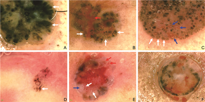
目的 探讨皮肤基底细胞癌的皮肤镜表现并分析其诊断价值,以了解皮肤镜诊断基底细胞癌的准确性。 方法 选取2017年7月1日—2019年12月31日就诊于阜阳市人民医院皮肤科门诊临床诊断疑似基底细胞癌的62例患者作为研究对象,其中男性29例,女性33例,发病年龄为(70.34±4.41)岁。通过单反数码相机及皮肤镜对门诊患者皮损部位进行取像观察,并行病理活检。以病理检查结果为金标准,评价皮肤镜诊断基底细胞癌的准确度,对皮肤镜及组织病理分型诊断结果的一致性进行分析。 结果 62例临床诊断疑似基底细胞癌患者中47例皮肤镜与病理结果一致,均为基底细胞癌。皮肤镜的诊断灵敏度为95.92%,特异度为92.31%,总符合率为95.16%,误诊率为7.69%,漏诊率为4.08%,Youden指数为0.882,Kappa值为0.858,且与病理诊断的一致性较高(P=0.999)。基底细胞癌皮肤镜下相关指标中,溃疡、多发浅表糜烂、亮红白色无结构区、蓝灰色卵圆巢、叶状结构等皮肤镜特征相关征象阳性率差异有统计学意义(均P < 0.05)。 结论 皮肤镜检查对基底细胞癌的诊断与病理组织检查结果吻合度较高,具有无创、便捷、准确度高的特点,值得临床推广。 Abstract:Objective To explore the dermatoscogic manifestations of basal cell carcinoma of the skin and analyze its diagnostic value, in order to understand the accuracy of dermatoscope in the diagnosis of basal cell carcinoma. Methods A total of 62 patients with suspected basal cell carcinoma were selected from the Dermatology Clinic of Fuyang People ' s Hospital from July 1, 2017 to December 31, 2019, including 29 males and 33 females, with an onset age of (70.34±4.41) years. The skin lesions of outpatients were observed by digital camera and dermatoscope, and pathological biopsy was performed. The results of pathological examination were taken as the gold standard, the accuracy of dermatoscope in the diagnosis of basal cell carcinoma was evaluated, and the consistency of dermatoscope and histopathological classification was analyzed. Results Among the 62 patients with suspected basal cell carcinoma, 47 were diagnosed as basal cell carcinoma by dermatoscope and pathology. The diagnostic sensitivity of dermatoscope was 95.92%, the specificity was 92.31%, the total coincidence rate was 95.16%, the misdiagnosis rate was 7.69%, the missed diagnosis rate was 4.08%, the Youden index was 0.882, the Kappa value was 0.858, and the consistency with pathological diagnosis was high (P=0.999). Among the dermatoscopic related indexes of basal cell carcinoma, there were significant differences in the positive rates of dermatoscogic features such as ulcer, multiple superficial erosion, bright red and white unstructured area, blue-gray oval nest, leafy structure and so on (all P < 0.05). Conclusion The diagnosis of basal cell carcinoma by dermatoscope is highly consistent with the results of pathological examination, and it has the characteristics of non-invasive, convenient and high accuracy, which is worthy of clinical promotion. -
Key words:
- Basal cell carcinoma /
- Dermatoscope /
- Diagnostic value
-
表 1 基底细胞癌皮肤镜特征[例(%)]
Table 1. Dermatographic features of basal cell carcinoma [cases (%)]
皮肤镜征象 阳性表现 蓝灰色卵圆巢 36(76.60) 灰蓝色小球 42(89.36) 叶状结构 13(27.66) 轮辐状结构 6(12.77) 溃疡 35(74.47) 树枝状血管 30(63.83) 短细毛细血管扩张 17(36.17) 多发浅表糜烂 7(14.89) 聚集性小点 6(12.77) 白色条纹/蝶蛹样结构 7(14.89) 同心环状结构 4(8.51) 亮红白色无结构区 4(8.51) 表 2 BCC与非BCC皮肤镜征象阳性表现情况比较(例)
Table 2. Comparison of positive manifestations of BCC and non-BCC dermatoscopy signs (cases)
项目 BCC
(47例)非BCC
(15例)χ2值 P值 树枝状血管 30 13 1.819 0.177 蓝白幕 3 1 0.001 0.999 短细毛细血管扩张 17 6 0.071 0.789 聚集性小点 6 4 0.759 0.384 同心环状结构 4 5 3.823 0.051 溃疡 35 4 9.180 0.002 多发浅表溃疡 7 6 4.325 0.038 亮红白色无结构区 4 7 8.880 0.003 灰蓝色小球 42 12 0.249 0.618 蓝灰色卵圆巢 36 6 6.969 0.008 轮辐状结构 6 0 0.321a 叶状结构 13 0 0.027a 白色条纹/蝶蛹样结构 7 2 0.001 0.999 色素网 0 2 0.056a 污斑 11 6 1.574 0.210 注:a为采用Fisher精确检验。 表 3 皮肤镜诊断与病理诊断结果比较(例)
Table 3. Comparison of results of dermatoscopy diagnosis and pathological diagnosis (cases)
皮肤镜 病理诊断 阳性 阴性 阳性 47 1 阴性 2 12 -
[1] APALLA Z, LALLAS A, SOTIRIOU E, et al. Epidemiological trends in skin cancer[J]. Dermatol Pract Concept, 2017, 7(2): 1-6. [2] 冯林, 余音, 张颖, 等. 浅表型基底细胞癌34例临床表现、皮肤镜及组织病理特征分析[J]. 临床皮肤科杂志, 2020, 49(7): 394-397. doi: 10.16761/j.cnki.1000-4963.2020.07.004FENG L, YU Y, ZHANG Y, et al. Clinical, dermatoscopic and histopathological features of superficial basal cell carcinoma: an analysis of 34 cases[J]. Journal of Clinical Dermatology, 2020, 49(7): 394-397. doi: 10.16761/j.cnki.1000-4963.2020.07.004 [3] YÉLAMOS O, BRAUN R P, LIOPYRIS K, et al. Usefulness of dermoscopy to improve the clinical and histopathologic diagnosis of skin cancers[J]. J Am Acad Dermatol, 2019, 80(2): 365-377. doi: 10.1016/j.jaad.2018.07.072 [4] PERIS K, FARGNOLI M C, GARBE C, et al. Diagnosis and treatment of basal cell carcinoma: European consensus-based interdisciplinary guidelines[J]. Eur J Cancer, 2019, 118: 10-34. doi: 10.1016/j.ejca.2019.06.003 [5] 中国中西医结合学会皮肤性病专业委员会皮肤影像学组, 中国医疗保健国际交流促进会皮肤科分会皮肤影像学组, 中华医学会皮肤性病学分会皮肤病数字化诊断亚学组, 等. 中国基底细胞癌皮肤镜特征专家共识(2019)[J]. 中华皮肤科杂志, 2019, 52(6): 371-377. doi: 10.3760/cma.j.issn.0412-4030.2019.06.001Dermatography Group of Dermatology and Venereal Disease Professional Committee of Chinese Society of Integrated Traditional Chinese and Western Medicine, Dermatography Group of Dermatology Branch of China Association for the Promotion of International Health Care, Dermatology Digital Diagnosis Sub-group of Dermatology Branch of Chinese Medical Association, etc. China Expert Consensus on Dermatoscopy Characteristics of Basal Cell Carcinoma (2019)[J]. Chinese Journal of Dermatology, 2019, 52(6): 371-377. doi: 10.3760/cma.j.issn.0412-4030.2019.06.001 [6] LANG B M, BALERMPAS P, BAUER A, et al. S2k guidelines for cutaneous basal cell carcinoma-part 1: Epidemiology, genetics and diagnosis[J]. J Dtsch Dermatol Ges, 2019, 17(1): 94-103. [7] ASGARI M M, MOFFET H H, RAY G T, et al. Trends in basal cell carcinoma incidence and identification of high-risk subgroups, 1998-2012[J]. JAMA Dermatol, 2015, 151(9): 976-981. doi: 10.1001/jamadermatol.2015.1188 [8] 刘子莲, 张倩, 吴雯婷, 等. 518例恶性皮肤肿瘤及癌前病变的回顾性分析[J]. 首都医科大学学报, 2018, 39(4): 602-606. doi: 10.3969/j.issn.1006-7795.2018.04.023LIU Z L, ZHANG Q, WU W T, etal. Skin cancer and precancerous skin lesions: retrospective analysis of 518 cases[J]. Journal of Capital Medical University, 2018, 39(4): 602-606. doi: 10.3969/j.issn.1006-7795.2018.04.023 [9] SÁNCHEZ G, NOVA J, RODRIGUEZ-HERNANDEZ A E, et al. Sun protection for preventing basal cell and squamous cell skin cancers[J]. Cochrane Database Syst Rev, 2016, 7(7): CD011161. DOI: 10.1002/14651858.CD011161.pub2. [10] CORONA R, DOGLIOTTI E, D ERRICO M, et al. Risk factors for basal cell carcinoma in a mediterranean population: Role of recreational sun exposure early in life[J]. Arch Dermatol, 2001, 137(9): 1162-1168. [11] WOLNER Z J, BAJAJ S, FLORES E, et al. Variation in dermoscopic features of basal cell carcinoma as a function of anatomical location and pigmentation status[J]. Br J Dermatol, 2018, 178(2): e136-e137. doi: 10.1111/bjd.15964 [12] 李薇薇, 涂平, 杨淑霞, 等. 皮肤镜对基底细胞癌鉴别诊断价值的初步研究[J]. 中华皮肤科杂志, 2013, 46(7): 480-484. doi: 10.3760/cma.j.issn.0412-4030.2013.07.009LI W W, TU P, YANG S X, et al. Dermoscopy in the differential diagnosis of basal cell carcinoma: a preliminary study[J]. Chinese Journal of Dermatology, 2013, 46(7): 480-484. doi: 10.3760/cma.j.issn.0412-4030.2013.07.009 [13] 张静. 基底细胞癌的皮肤镜表现与病理类型的相关性研究[J]. 世界最新医学信息文摘, 2019, 19(23): 20-21. https://www.cnki.com.cn/Article/CJFDTOTAL-WMIA201923010.htmZHANG J. Study on the correlation between cutaneous manifestations and pathological types of basal cell carcinoma[J]. World Latest Medical Information Digest, 2019, 19(23): 20-21. https://www.cnki.com.cn/Article/CJFDTOTAL-WMIA201923010.htm [14] REITER O, MIMOUNI I, GDALEVICH M, et al. The diagnostic accuracy of dermoscopy for basal cell carcinoma: A systematic review and meta-analysis[J]. J Am Acad Dermatol, 2019, 80(5): 1380-1388. doi: 10.1016/j.jaad.2018.12.026 [15] MCKEE P H, CALONJE E, GRANTER S R, 等. 皮肤病理学: 与临床的联系[M]. 朱学骏, 孙建方, 译. 4版. 北京: 北京大学医学出版社, 2017: 1167-1184.MCKEE P H, CALONJE E, GRANTER S R, et al. Dermatopathology: the relationship with clinic[M]. ZHU X J, SUN J F, translated. 4th ed. Beijing: Peking University Medical Press, 2017: 1167-1184. [16] LOMBARDI M, PAMPENA R, BORSARI S, et al. Dermoscopic features of basal cell carcinoma on the lower limbs: A chameleon![J]. Dermatology, 2017, 233(6): 482-488. doi: 10.1159/000487300 -


 下载:
下载:


