

IL-33/ST2 pathway in atherosclerosis: the IL-33 paradox
-
摘要:
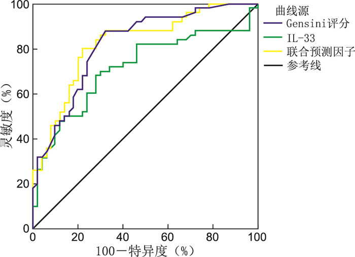
目的 探讨人和动物实验模型中IL-33/可溶性生长刺激表达基因2蛋白(sST2)通路与冠状动脉硬化之间的关系。 方法 纳入2019年1—12月就诊于新疆维吾尔自治区人民医院心内科明确诊断为急性心肌梗死的患者(AMI组,50例)和稳定型冠心病患者(CAD组,50例)。收集研究对象冠状动脉血标本,用ELISA法检测IL-33水平。动物实验模型中,收集ApoE基因敲除小鼠的下腔静脉血液,检测TG、总胆固醇(CHOL) 浓度,用ELISA法计算样品的凝血酶-抗凝血酶复合物(TAT)水平。 结果 AMI组患者的IL-33、Gensini评分显著高于CAD组(均P<0.05)。Pearson相关性分析显示, Gensini评分与IL-33表达水平呈正相关关系(P<0.05)。多元线性回归分析显示,IL-33(β=0.295,P=0.001)水平是Gensini评分的独立影响因素。ROC曲线分析可知,IL-33、Gensini评分及其联合预测的AUC分别为0.716、0.820、0.864。动物实验模型中,不同浓度IL-33干预后ApoE基因敲除的小鼠TAT浓度、TG、CHOL浓度随着干预时间的延长而降低(均P<0.05)。 结论 AMI患者冠脉血IL-33通过IL-33/ST2依赖性途径促进动脉硬化的进展,并诱导血栓形成。IL-33可降低动脉粥样硬化的ApoE小鼠的血脂来抑制动脉粥样硬化的进展。 -
关键词:
- 白细胞介素-33 /
- 可溶性生长刺激表达基因2蛋白 /
- 动脉硬化
Abstract:Objective This study aimed to investigate the relationship between the IL-33/soluble growth stimulation expressed gene 2 (ST2) pathway and coronary arteriosclerosis in human and animal models. Methods The clinical trial included patients with a clear diagnosis of acute myocardial infarction (AMI group, n=50) and stable coronary artery disease (CAD group, n=50) who visited the Department of Cardiology, People ' s Hospital of Xinjiang Uygur Autonomous Region from January 2019 to December 2019. Coronary blood specimens were collected from the study subjects, and IL-33 levels were detected by ELISA. In the animal experimental model, blood was collected from inferior vena cava of ApoE knockout mice, TG and cholesterol (CHOL) concentrations were measured. In addition, the thrombin-antithrombin complex (TAT) levels of the samples were calculated by ELISA. Results IL-33 and Gensini scores in AMI group were significantly higher than those in CAD group (all P < 0.05). Pearson correlation analysis showed a positive correlation between Gensini score and IL-33 level expression (P < 0.05). Multiple linear regression analysis showed that Gensini score levels were independently correlated with IL-33 (β=0.295, P=0.001) levels. The area under the curve (AUC) of the Gensini score, the AUC for IL-33, Gensini score and joint predictors were 0.716, 0.820 and 0.864, respectively. In the animal experimental model, the TAT concentration, TG and CHOL concentrations in ApoE knockout mice decreased with the increase of intervention time after different concentrations of IL-33 intervention (all P < 0.05). Conclusion Coronary blood IL-33 in patients with AMI accelerates the progression of atherosclerosis through the IL-33/ST2 pathway and induces thrombosis. IL-33 reduces lipids in atherosclerotic ApoE mice to inhibit the progression of atherosclerosis. -
表 1 AMI组和CAD组患者一般临床资料比较
Table 1. Comparison of general clinical data between patients in AMI group and CAD group
指标 AMI组(n=50) CAD组(n=50) 统计量 P值 基线资料 年龄(x±s,岁) 57.6±12.3 60.9±7.9 1.617a 0.109 性别(男/女,例) 38/12 34/16 0.794b 0.373 BMI(x±s) 26.6±6.9 24.7±3.8 1.613a 0.110 吸烟[例(%)] 24(48.0) 28(56.0) 0.641b 0.423 收缩压(x±s,mm Hg) 129.4±23.1 136.9±20.2 1.754a 0.083 舒张压(x±s,mm Hg) 76.9±13.8 78.4±11.8 0.583a 0.561 既往病史 高血压[例(%)] 29(58.0) 23(46.0) 1.442b 0.230 糖尿病[例(%)] 33(66.0) 37(74.0) 0.762b 0.383 高脂血症[例(%)] 43(86.0) 41(82.0) 0.298b 0.585 生化指标 TG(x±s,mmol/L) 1.5±0.8 1.7±1.4 0.772a 0.442 CHOL(x±s,mmol/L) 4.4±1.2 4.2±1.3 0.614a 0.054 HDL-C(x±s,mmol/L) 1.0±0.3 0.9±0.2 2.005a 0.061 LDL-C(x±s,mmol/L) 3.0±1.0 2.3±0.9 3.603a 0.001 LPa[M(P25, P75),mmol/L] 186.7(117.7, 380.0) 228.8(134.0, 349.0) 0.411c 0.682 C-反应蛋白[M(P25, P75), μg/L] 3.8(2.5, 12.3) 2.5(1.7, 3.5) -1.781c 0.079 血肌酐[M(P25, P75),μg/L] 69.5(57.9, 78.5) 62.5(55.5, 72.6) -1.557c 0.123 NT-proBNP[M(P25, P75), pmol/L] 238.6(97.1, 573.5) 29.0(15.7, 64.0) 0.062c 0.066 注:1 mm Hg =0.133 kPa,a为t值,b为χ2值,c为Z值。  下载: 导出CSV
表 2 AMI组和CAD组患者IL-33水平和Gensini评分比较(x ±s)
Table 2. The levels of IL-33 and Gensini score in patients with AMI andCAD(x ±s)
组别 例数 IL-33(pg/mL) Gensini评分(分) AMI 50 139.1±43.9 70.6±36.9 CAD 77 111.9±28.9 32.2±15.7 t值 3.656 6.040 P值 <0.001 <0.001
下载: 导出CSV
表 3 临床指标与Gensini评分的相关性分析
Table 3. Correlation between Gensini score and clinical index
项目 r值 P值 IL-33(pg/mL) 0.314 0.001 LDL-C(mmol/L) 0.113 0.270
下载: 导出CSV
表 4 相关临床指标与Gensini评分的多元线性回归分析
Table 4. Multiple linear regression analysis of Gensini score and clinical indexes
变量 β SE B t值 P值 IL-33(pg/mL) 0.295 0.090 0.314 3.270 0.001 LDL-C(mmol/L) 4.003 3.606 0.113 1.110 0.270 注:R2=0.607,调整R2=0.565,F=1.736,P=0.004。连续变量均以实际值赋值。
下载: 导出CSV
表 5 不同浓度的IL-33干预时的CHOL浓度随时间变化(x ±s)
Table 5. Changes in CHOL concentration over time during intervention with different concentrations of IL-33 (x ±s)
组别 例数 4周 6周 8周 F值 P值 对照组 7.25±1.14 6.90±0.96 7.02±1.04 0.144 0.867 IL-33(1.0×10⁶ pg/mL) 5.96±0.98 6.02±0.81 5.37±0.85a 0.841 0.455 IL-33(2.5×10⁶ pg/mL) 5.45±0.99a 4.88±1.14a 4.59±0.99a 0.882 0.439 IL-33(5.0×10⁶ pg/mL) 5.70±0.75a 5.44±0.88a 4.69±0.66a 2.382 0.135 F值 3.384 4.088 7.895 P值 0.044 0.025 0.002 注:与同一时间对照组比较,aP<0.05。
下载: 导出CSV
表 6 不同浓度的IL-33干预时的TG浓度随时间变化(x ±s)
Table 6. Changes in TG concentration over time during intervention with different concentrations of IL-33 (x ±s)
组别 例数 4周 6周 8周 F值 P值 对照组 4.34±0.62 4.45±0.56 4.30±0.64 0.085 0.919 IL-33(1.0×10⁶ pg/mL) 3.99±0.44 3.63±0.41 3.88±0.38a 1.054 0.379 IL-33(2.5×10⁶ pg/mL) 3.93±0.57 3.37±0.66a 3.27±0.41a 2.094 0.166 IL-33(5.0×10⁶ pg/mL) 3.82±0.74 3.63±0.46 3.31±0.35a 2.382 0.358 F值 0.694 3.962 5.883 P值 0.569 0.027 0.007 注:与同一时间对照组比较,aP<0.05。
下载: 导出CSV
表 7 不同浓度的IL-33干预时的TAT浓度随时间变化(x ±s)
Table 7. Changes in TAT concentration over time during intervention with different concentrations of IL-33 (x ±s)
组别 4周 6周 8周 F值 P值 对照组 0.95±0.08 0.95±0.06 0.92±0.07 0.165 0.850 IL-33(1.0×10⁶ pg/mL) 0.85±0.10 0.74±0.06a 0.73±0.10ac 2.961 0.900 IL-33(2.5×10⁶ pg/mL) 0.79±0.07a 0.70±0.07a 0.62±0.10ac 4.886 0.028 IL-33(5.0×10⁶ pg/mL) 0.77±0.08a 0.65±0.05ab 0.60±0.08ac 7.287 0.008 F值 4.630 21.431 12.940 P值 0.016 <0.001 <0.001 注:与同一时间对照组比较,aP<0.05;与同一浓度4周和6周比较,bP<0.05;与同一浓度4周和8周比较,cP<0.05;与同一浓度6周和8周比较,dP<0.05。
下载: 导出CSV
-
[1] PROUT M S, KYLE R L, RONCHESE F, et al. IL-4 Is a key requirement for IL-4- and IL-4/IL-13-expressing CD4 Th2 subsets in lung and skin[J]. Frontiers in immunology, 2018, 9: 1211. DOI: 10.3389/fimmu.2018.01211. [2] CAYROL C, GIRARD J P. Interleukin-33 (IL-33): a nuclear cytokine from the IL-1 family[J]. Immunological reviews, 2018, 281(1): 154-168. doi: 10.1111/imr.12619 [3] WOLF D, LEY K. Immunity and inflammation in atherosclerosis[J]. Circ Res, 2019, 124(2): 315-327. doi: 10.1161/CIRCRESAHA.118.313591 [4] LUNDBECH M, KRAG A E, CHRISTENSEN T D, et al. Thrombin generation, thrombin-antithrombin complex, and prothrombin fragment F1+2 as biomarkers for hypercoagulability in cancer patients[J]. Thromb Res, 2020, 186: 80-85. doi: 10.1016/j.thromres.2019.12.018 [5] BRETSCHER P. On analyzing how the Th1/Th2 phenotype of an immune response is determined: classical observations must not be ignored[J]. Front Immunol, 2019, 10: 1234. DOI: 10.3389/fimmu.2019.01234. [6] STARK J M, TIBBITT C A, COQUET J M. The metabolic requirements of Th2 cell differentiation[J]. Front Immunol, 2019, 10: 2318. DOI: 10.3389/fimmu.2019.02318. [7] MURDACA G, GRECO M, TONACCI A, et al. IL-33/IL-31 axis in immune-mediated and allergic diseases[J]. Int J Mol Sci, 2019, 20(23): 5856. DOI: 10.3390/ijms20235856. [8] ITO T, KAGEYAMA R, NAKAZAWA S, et al. Understanding the significance of cytokines and chemokines in the pathogenesis of alopecia areata[J]. Exp Dermatol, 2020, 29(8): 726-732. doi: 10.1111/exd.14129 [9] LI W P, LI Y Y, JIN J. The essential function of IL-33 in metabolic regulation[J]. Acta Biochim Biophys Sin (Shanghai), 2020, 52(7): 768-775. doi: 10.1093/abbs/gmaa045 [10] RUTERBUSCH M, PRUNER K B, SHEHATA L, et al. In vivo CD4+ T cell differentiation and function: revisiting the Th1/Th2 paradigm[J]. Annu Rev Immunol, 2020, 38: 705-725. doi: 10.1146/annurev-immunol-103019-085803 [11] 中华医学会, 中华医学会杂志社, 中华医学会全科医学分会, 等. 稳定性冠心病基层诊疗指南(2020年)[J]. 中华全科医师杂志, 2021, 20(3): 265-273.Chinese Medical Association, Chinese Medical Association magazine, Chinese Medical Association Branch of General Practice, etc. Primary Diagnosis and Treatment Guidelines for Stable Coronary Heart Disease (2020)[J]. Chinese Journal of General Practice, 2021, 20(3): 265-273. [12] DEMYANETS S, STOJKOVIC S, HUBER K, et al. The paradigm change of IL-33 in vascular biology[J]. Int J Mol Sci, 2021, 22(24): 13288. DOI: 10.3390/ijms222413288. [13] PUSCEDDU I, DIEPLINGER B, MUELLER T. ST2 and the ST2/IL-33 signalling pathway-biochemistry and pathophysiology in animal models and humans[J]. Clinica Chimica Acta, 2019, 495: 493-500. doi: 10.1016/j.cca.2019.05.023 [14] PFEILER S, WINKELS H, KELM M, et al. IL-1 family cytokines in cardiovascular disease[J]. Cytokine, 2019, 122: 154215. DOI: 10.1016/j.cyto.2017.11.009. [15] AJITHKUMAR V, AXEL K. Interleukin (IL)-33 and the IL-1 family of cytokines-regulators of inflammation and tissue homeostasis[J]. Cold Spring Harb Perspect Biol, 2019, 11(3): a028506. DOI: 10.1101/cshperspect.a028506. [16] KONG D H, KIM Y K, KIM M R, et al. Emerging roles of vascular cell adhesion molecule-1 (VCAM-1) in immunological disorders and cancer[J]. Int J Mol Sci, 2018, 19(4): 1057. DOI: 10.3390/ijms19041057. [17] LIU X X, XIAO Y C, PAN Y, et al. The role of the IL-33/ST2 axis in autoimmune disorders: friend or foe?[J]. Cytokine Growth Factor Rev, 2019, 50: 60-74. doi: 10.1016/j.cytogfr.2019.04.004 [18] LI J, MENG Q H, FU J, et al. Novel insights: Dynamic foam cells derived from the macrophage in atherosclerosis[J]. J Cell Physiol, 2021, 236(9): 6154-6167. doi: 10.1002/jcp.30300 [19] BÄCK M, JR A Y, TABAS I, et al. Inflammation and its resolution in atherosclerosis: mediators and therapeutic opportunities[J]. Nat Rev Cardiol, 2019, 16(7): 389-406. [20] XIE Z L, WANG X D, LIU X X, et al. Adipose-derived exosomes exert proatherogenic effects by regulating macrophage foam cell formation and polarization[J]. J Am Heart Assoc, 2018, 7(5): e007442. DOI: 10.1161/JAHA.117.007442. [21] CAYROL C. IL-33, an alarmin of the IL-1 family involved in allergic and non allergic inflammation: focus on the mechanisms of regulation of its activity[J]. Cells, 2021, 11(1): 107. doi: 10.3390/cells11010107 [22] TANG H N, LIU N, FENG X J, et al. Circulating levels of IL-33 are elevated by obesity and positively correlated with metabolic disorders in Chinese adults[J]. J Transl Med, 2021, 19(1): 52. DOI: 10.1186/s12967-021-02711-x. [23] AOKI S, HAYAKAWA M, OZAKI H, et al. ST2 gene expression is proliferation-dependent and its ligand, IL-33, induces inflammatory reaction in endothelial cells[J]. Mol Cell Biochem, 2010, 335(1-2): 75-81. doi: 10.1007/s11010-009-0244-9 [24] MCLAREN J E, MICHAEL D R, SALTER R C, et al. IL-33 reduces macrophage foam cell formation[J]. J Immunol, 2010, 185(2): 1222-1229. doi: 10.4049/jimmunol.1000520 [25] DEMYANETS S, KONYA V, KASTL S P, et al. Interleukin-33 induces expression of adhesion molecules and inflammatory activation in human endothelial cells and in human atherosclerotic plaques[J]. Arterioscler Thromb Vasc Biol, 2011, 31(9): 2080-2089. doi: 10.1161/ATVBAHA.111.231431 [26] WU F Q, HE M A, WEN Q, et al. Associations between variants in IL-33/ST2 signaling pathway genes and coronary heart disease risk[J]. Int J Mol Sci, 2014, 15(12): 23227-23239. doi: 10.3390/ijms151223227 -

 点击查看大图
计量
- 文章访问数: 280
- HTML全文浏览量: 51
- PDF下载量: 14
- 被引次数: 0
