

Feasibility study of DCE-MRI parameters combined with CA199, CA125, HE4 in differentiating BOT from stage Ⅰ EOC
-
摘要:
目的 明确诊断卵巢交界性肿瘤(BOT)、Ⅰ期上皮性卵巢癌(EOC),对临床选择治疗方式有关键意义。本研究以动态增强磁共振(DCE-MRI)参数结合糖类抗原199(CA199)、CA125、(HE4)人附睾蛋白4鉴别诊断BOT与Ⅰ期EOC,分析其临床应用的可行性。 方法 收集2018年1月—2022年4月阜阳市肿瘤医院收治BOT患者95例(BOT组),Ⅰ期EOC患者62例(EOC组)。对比2组DCE-MRI征象、定量参数及CA199、CA125、HE4表达水平,并分析不同检查对BOT与Ⅰ期EOC的诊断效果。 结果 2组常规形态学比较差异无统计学意义(均P>0.05);EOC组达峰时间(TTP)值低于BOT组,对比剂最大浓度(Cmax)、AUC、最大线性斜率(MS)值均高于BOT组(均P<0.05)。EOC组CA125、HE4、CA199表达水平均高于BOT组(均P<0.05)。ROC曲线结果显示:DCE-MRI参数(TTP、Cmax、AUC、MS)和CA199、CA125、HE4对BOT与Ⅰ期EOC鉴别曲线下面积(AUC)分别为0.789、0.682、0.799、0.786、0.792、0.789、0.816,联合诊断的AUC值为0.956,高于四者单独诊断(P<0.05),联合诊断灵敏度、特异度分别为97.60%、94.80%。 结论 BOT与Ⅰ期EOC在DCE-MRI参数及CA199、CA125、HE4表达水平中存在差异,DCE-MRI联合血清学指标对两者鉴别诊断可行性高。 Abstract:Objective Diagnosis of borderline ovarian tumor (BOT) and stage Ⅰ epithelial ovarian cancer (EOC) is of key significance for clinical selection of treatment. In this study, dynamic enhanced MRI (DCE-MRI) parameters combined with glycoantigen 199 (CA199), CA125, and human epididymal protein 4 (HE4) were used to differentially diagnose BOT and stage Ⅰ EOC, and analyze the feasibility of the clinical application. Methods A total of 95 BOT patients admitted to Fuyang Cancer Hospital from January 2018 to April 2022 were collected as BOT group, and 62 stage Ⅰ EOC patients were collected as EOC group. The DCE-MRI signs, quantitative parameters, and expression levels of CA199, CA125, and HE4 were compared between the two groups, and the diagnostic effects of different examinations on BOT and stage Ⅰ EOC were analyzed. Results There was no difference in conventional morphology between the two groups (all P > 0.05). The time to peak (TTP) values of EOC group were lower than those of BOT group, while the contrast agent maximum concentration (Cmax), area under curve (AUC) and maximum linear slope (MS) values of EOC group were higher than those of BOT group (all P < 0.05). Expression levels of CA125, HE4 and CA199 in EOC group were higher than those in BOT group (all P < 0.05). ROC curve results show DCE-MRI parameters (TTP, Cmax, AUC, MS) combined with CA199, CA125, HE4 for BOT and Stage Ⅰ EOC were 0.789, 0.682, 0.799, 0.786, 0.792, 0.789, 0.816, respectively. The AUC value of combined diagnosis was 0.956, which was higher than that of the four patients diagnosed alone (P < 0.05). The sensitivity and specificity of combined diagnosis were 97.60% and 94.80%, respectively. Conclusion There are differences in DCE-MRI parameters and CA199, CA125, HE4 expression levels between BOT and stage Ⅰ EOC, and DCE-MRI combined with serological indicators is highly feasible for the differential diagnosis of the two. -

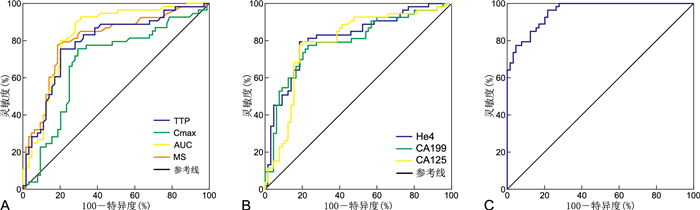
图 1 DCE-MRI结合CA199、CA125、HE4对BOT与Ⅰ期EOC鉴别的ROC曲线
注:A为TTP、Cmax、AUC、MS对BOT与Ⅰ期EOC鉴别ROC曲线;B为CA199、CA125、HE4对BOT与Ⅰ期EOC鉴别ROC曲线;C为联合诊断对BOT与Ⅰ期EOC鉴别ROC曲线。
Figure 1. ROC curve of DCE-MRI combined with CA199, CA125 and HE4 for BOT and Stage Ⅰ EOC identification

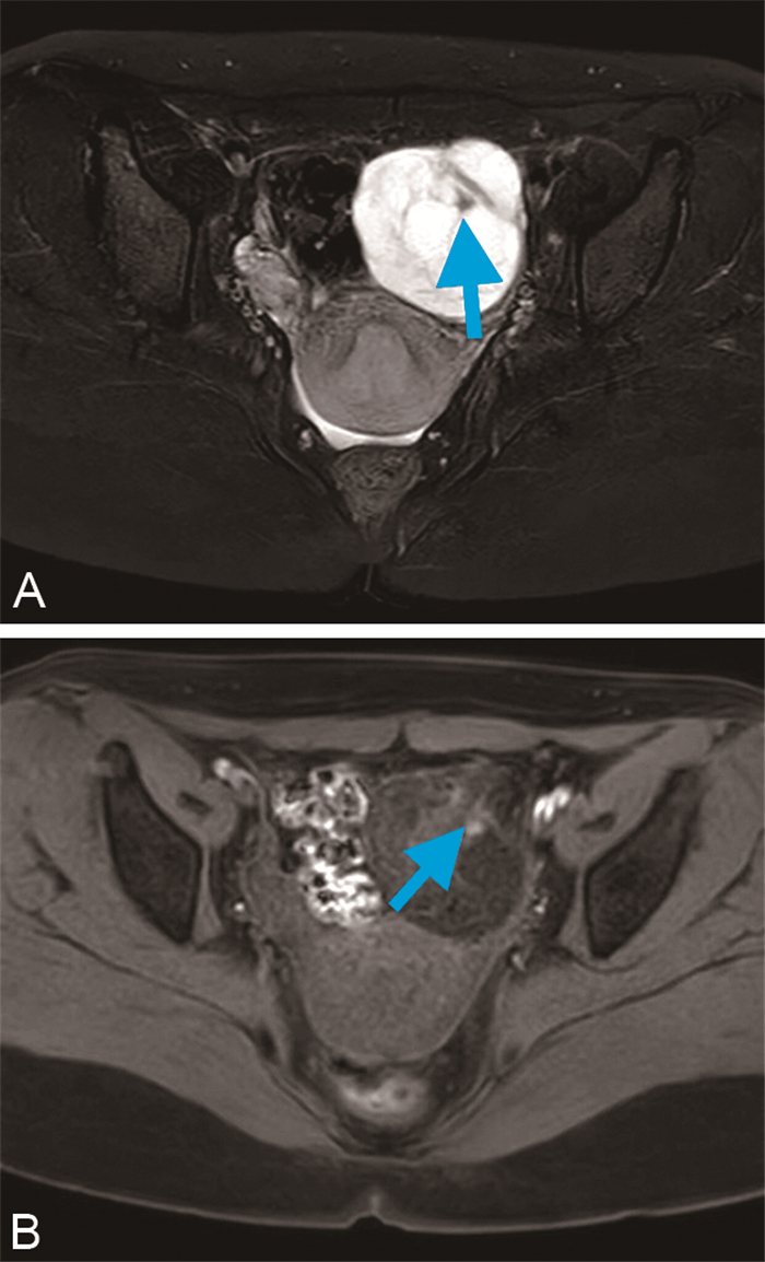
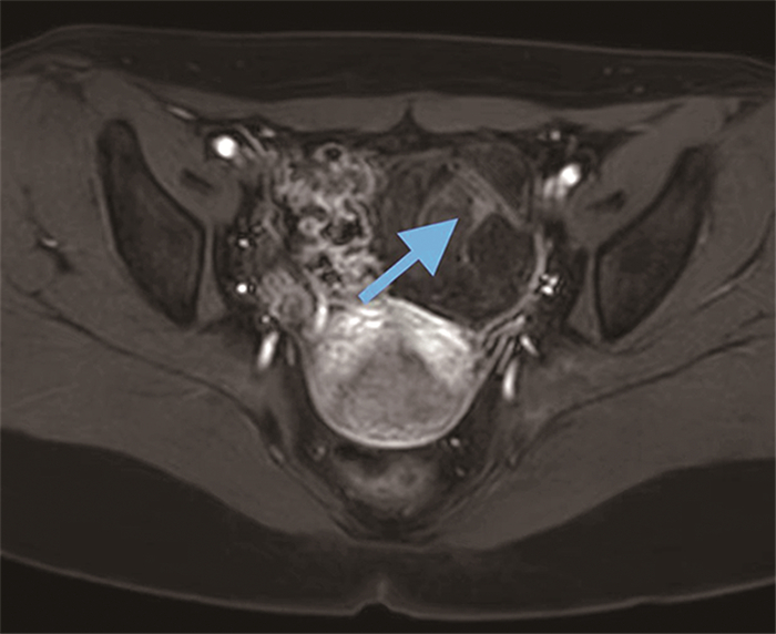
图 3 EOC组典型病例图像
注:盆腔内双侧附件区囊实性包块(箭头)。
Figure 3. Images of typical cases in the EOC group
表 1 2组卵巢肿瘤患者影像学征象比较[例(%)]
Table 1. Comparison of imaging findings of ovarian tumor patients in two groups[cases (%)]
组别 例数 肿瘤类型 强化程度 病灶部位 分隔数 囊性 实性 囊实性 轻 中 重 单侧 双侧 0~4 ≥5 BOT组 95 44(46.31) 22(23.16) 29(30.53) 26(27.37) 40(42.10) 29(30.53) 81(85.26) 14(14.74) 33(34.74) 62(65.26) EOC组 62 19(30.65) 14(22.58) 29(46.77) 15(24.19) 25(40.32) 22(35.48) 46(74.19) 16(25.81) 24(38.81) 38(61.29) 统计量 4.982a -0.656b 2.297a 0.256a P值 0.082 0.512 0.084 0.612 注:a为χ2值,b为Z值。  下载: 导出CSV
表 2 2组卵巢肿瘤患者DCE-MRI定量参数比较(x±s)
Table 2. Comparison of DCE-MRI quantitative parameters in two groups of ovarian tumor patients(x±s)
组别 例数 TTP (s) Cmax (mmol/L) AUC MS BOT组 95 3.81±0.36 0.14±0.05 0.52±0.10 0.20±0.02 EOC组 62 3.45±0.31 0.20±0.06 0.67±0.11 0.37±0.07 t值 6.462 6.785 8.829 22.347 P值 <0.001 <0.001 <0.001 <0.001
下载: 导出CSV
表 3 2组卵巢肿瘤患者CA199、CA125、HE4表达水平比较(x±s)
Table 3. Comparison of expression levels of CA199, CA125 and HE4 in two groups of ovarian tumor patients(x±s)
组别 例数 CA199 (U/mL) CA125 (U/mL) HE4 (pmol/L) BOT组 95 62.45±7.15 19.63±4.15 83.55±7.18 EOC组 62 88.79±9.47 315.27±14.60 167.15±10.33 t值 19.814 186.439 59.825 P值 <0.001 <0.001 <0.001
下载: 导出CSV
表 4 联合诊断对BOT与Ⅰ期EOC的鉴别效果
Table 4. Effect of combined diagnosis in differentiating BOT from Stage Ⅰ EOC
项目 截断值 AUC 95% CI 灵敏度 特异度 P值 TTP 3.19 0.789 0.704~0.873 0.744 0.628 <0.001 Cmax 0.19 0.682 0.581~0.782 0.636 0.669 <0.001 AUC 0.63 0.799 0.754~0.906 0.726 0.652 <0.001 MS 0.18 0.786 0.722~0.887 0.751 0.730 <0.001 CA199 76.26 0.792 0.707~0.877 0.638 0.776 <0.001 CA125 220.74 0.789 0.697~0.877 0.625 0.722 <0.001 HE4 116.55 0.816 0.734~0.884 0.811 0.732 <0.001 联合 0.956 0.925~0.986 0.976 0.948 <0.001
下载: 导出CSV
-
[1] 刘文霞, 刘利, 贡琦, 等. CT, 动态增强磁共振成像及弥散加权成像对卵巢囊腺癌与囊腺瘤的鉴别效果分析[J]. 中国医学装备, 2022, 19(9): 45-49. https://www.cnki.com.cn/Article/CJFDTOTAL-YXZB202209011.htmLIU W X, LIU L, Gong Q, et al. Effectiveness of CT, dynamic enhanced magnetic resonance imaging and diffusion-weighted imaging in differentiating ovarian cystic adenocarcinoma from cystic adenoma[J]. China Medical Equipment, 2022, 19(9): 45-49. https://www.cnki.com.cn/Article/CJFDTOTAL-YXZB202209011.htm [2] 张俊梅, 刘洁梅, 李琳可, 等. MRI联合血清肿瘤标志物对BOT及Ⅰ期EOC的鉴别诊断价值[J]. 中国CT和MRI杂志, 2022, 20(8): 177-179. https://www.cnki.com.cn/Article/CJFDTOTAL-CTMR202208049.htmZHANG J M, LIU J M, Li LK, et al. Differential diagnostic value of MRI combined with serum tumor markers for BOT and stage Ⅰ EOC[J]. Chinese Journal of CT and MRI, 2022, 20(8): 177-179. https://www.cnki.com.cn/Article/CJFDTOTAL-CTMR202208049.htm [3] 刘格格, 王丽华, 李燕华. 上皮性卵巢癌患者的BRCA1/2基因状态与其临床病理特征的相关性分析[J]. 中华全科医学, 2023, 21(5): 749-752, 768. doi: 10.16766/j.cnki.issn.1674-4152.002974LIU G G, WANG L H, LI Y H. Correlation analysis of BRCA1/2 gene status and its clinicopathological features in patients with epithelial ovarian cancer[J]. Chinese Journal of General Practice, 2023, 21(5): 749-752, 768. doi: 10.16766/j.cnki.issn.1674-4152.002974 [4] 王艳, 曲源, 陈杰, 等. 动态增强成像影像组学分析鉴别良恶性腮腺病变的价值研究[J]. 中华全科医学, 2023, 21(3): 463-468.WANG Y, QU Y, CHEN J, TIAN H, Shang Yurong. Study on the value of dynamic enhanced imaging histological analysis to identify benign and malignant parotid lesions[J]. Chinese Journal of General Practice, 2023, 21(3): 463-468. [5] 李向荣, 杜旋, 杨娟. 血清CA199、CA125、HE4联合检测在卵巢癌诊断中的应用价值[J]. 实用癌症杂志, 2020, 35(6): 1015-1018. doi: 10.3969/j.issn.1001-5930.2020.06.039LI X R, DU X, YANG J. The value of combined serum CA199, CA125 and HE4 in the diagnosis of ovarian cancer[J]. The Practical Journal of Cancer, 2020, 35(6): 1015-1018. doi: 10.3969/j.issn.1001-5930.2020.06.039 [6] 张晖, 连鹏, 杨璐, 等. DCE-MRI半定量参数联合血清学指标鉴别卵巢交界性肿瘤和上皮性卵巢癌的价值[J]. 重庆医学, 2021, 50(14): 2402-2406, 2411. doi: 10.3969/j.issn.1671-8348.2021.14.014ZHANG F, LIAN P, YANG L, et al. The value of DCE-MRI semi-quantitative parameters combined with serological indices to identify ovarian junctional tumors and epithelial ovarian cancer[J]. Chongqing Medicine, 2021, 50(14): 2402-2406, 2411. doi: 10.3969/j.issn.1671-8348.2021.14.014 [7] 李青, 赵玉娇, 黄黎香, 等. MRI磁共振扫描仪对卵巢交界性肿瘤和上皮性卵巢癌的鉴别诊断价值[J]. 中国医疗器械信息, 2020, 26(23): 41-42. doi: 10.3969/j.issn.1006-6586.2020.23.018LI Q, ZhHAN Y J, HUANG L X, et al. Differential diagnostic value of MRI magnetic resonance scanner between ovarian junctional tumor and epithelial ovarian cancer[J]. China Medical Device Information, 2020, 26(23): 41-42. doi: 10.3969/j.issn.1006-6586.2020.23.018 [8] 毛咪咪, 冯峰, 李海明, 等. 定量动态增强MRI在鉴别交界性与恶性上皮性卵巢肿瘤中的价值[J]. 临床放射学杂志, 2019, 38(4): 669-674. doi: 10.13437/j.cnki.jcr.2019.04.023MAO M M, FENG F, LI H M, et al. Value of quantitative dynamic-enhanced MRI in differentiating junctional from malignant epithelial ovarian tumors[J]. Journal of Clinical Radiology, 2019, 38(4): 669-674. doi: 10.13437/j.cnki.jcr.2019.04.023 [9] 安永玉, 李文华. DWI及常规MRI在卵巢交界性肿瘤与上皮性卵巢癌中的鉴别诊断价值[J]. 医学影像学杂志, 2019, 29(2): 277-282. https://www.cnki.com.cn/Article/CJFDTOTAL-XYXZ201902036.htmAN Y Y, LI WH. Differential diagnostic value of DWI and conventional MRI in ovarian junctional tumors and epithelial ovarian cancer[J]. Journal of Medical Imaging, 2019, 29(2): 277-282. https://www.cnki.com.cn/Article/CJFDTOTAL-XYXZ201902036.htm [10] 韩旭, 孙美玉. CT及MRI在卵巢上皮交界性与恶性肿瘤鉴别中的研究进展[J]. 实用放射学杂志, 2019, 35(3): 154-159.HAN X, SuUN M Y. Research progress of CT and MRI in differentiating ovarian epithelial junction from malignant tumors[J]. Journal of practical radiology, 2019, 35(3): 154-159. [11] 左金, 闫海龙, 韩东明. DCE-MRI在卵巢交界性肿瘤与上皮性卵巢癌中的鉴别诊断价值[J]. 医学影像学杂志, 2020, 30(5): 798-802. https://www.cnki.com.cn/Article/CJFDTOTAL-XYXZ202005021.htmZUO J, YAN H L, HAN D M. Differential diagnostic value of DCE-MRI in ovarian junctional tumors and epithelial ovarian cancer[J]. Journal of Medical Imaging, 2020, 30(5): 798-802. https://www.cnki.com.cn/Article/CJFDTOTAL-XYXZ202005021.htm [12] 来金欣, 叶嘉琪, 陈杰荣, 等. 血清糖类抗原125联合人附睾蛋白4及糖类抗原199检测诊断上皮性卵巢癌的意义[J]. 中国妇幼保健, 2020, 35(24): 4674-4677. https://www.cnki.com.cn/Article/CJFDTOTAL-ZFYB202024009.htmLAI J X, CHEN J R, Gan HCQ, et al. Diagnosis of epithelial ovarian cancer by serum glycoantigen 125 in combination with human epithelial protein 4 and glycoantigen 199[J]. China Maternal and Child Health, 2020, 35(24): 4674-4677. https://www.cnki.com.cn/Article/CJFDTOTAL-ZFYB202024009.htm [13] 胡原, 单秀红. MRI图像特征结合ADC值在鉴别卵巢交界性囊腺瘤与囊腺癌中的应用[J]. 中国CT和MRI杂志, 2020, 18(8): 129-131. https://www.cnki.com.cn/Article/CJFDTOTAL-CTMR202008040.htmHU Y, SHAN X H. MRI image features combined with ADC values in differentiating ovarian junctional cystadenoma from cystic adenocarcinoma[J]. Chinese Journal of CT and MRI, 2020, 18(8): 129-131. https://www.cnki.com.cn/Article/CJFDTOTAL-CTMR202008040.htm [14] 刘碧英, 张小镇, 何岩燕, 等. DCE-MRI定量分析、多b值DWI联合应用对卵巢交界性、恶性肿瘤的鉴别价值及与Ki-67的相关性研究[J]. 临床放射学杂志, 2021, 40(4): 757-762. https://www.cnki.com.cn/Article/CJFDTOTAL-LCFS202104029.htmLIU B Y, ZHABF Z, HE Y Y, et al. The value of DCE-MRI quantitative analysis and multi-b value DWI for the differentiation of ovarian junctional and malignant tumors and the correlation with Ki-67[J]. Journal of Clinical Radiology, 2021, 40(4): 757-762. https://www.cnki.com.cn/Article/CJFDTOTAL-LCFS202104029.htm [15] 钱晨. CA125, CA199联合HE4在卵巢癌患者中表达及诊断效果研究[J]. 结直肠肛门外科, 2021, 27(1): 87-88. https://www.cnki.com.cn/Article/CJFDTOTAL-SWCX202316014.htmQIAN C. Study on the expression and diagnostic effect of CA125, CA199 combined with HE4 in patients with ovarian cancer[J]. Colorectal Surgery, 2021, 27(1): 87-88. https://www.cnki.com.cn/Article/CJFDTOTAL-SWCX202316014.htm [16] 蒋东葵, 陈兆亚, 马冬梅, 等. 血清胃泌素释放肽前体、甲胎蛋白和糖类抗原199单项及联合检测在卵巢癌诊断中的价值[J]. 诊断学理论与实践, 2019, 18(2): 209-214. https://www.cnki.com.cn/Article/CJFDTOTAL-ZDLS201902022.htmJIANG D K, CHEN Z Y, MA D M, et al. Value of serum gastrin-releasing peptide precursors, methemoglobin and glycoantigen 199 alone and in combination in the diagnosis of ovarian cancer[J]. Diagnostics Theory and Practice, 2019, 18(2): 209-214. https://www.cnki.com.cn/Article/CJFDTOTAL-ZDLS201902022.htm [17] 韩梅, 马明杰, 连俊, 等. 血清AFP、CEA、CA199、CA125、HE4联合检测在卵巢癌诊断中的应用价值[J]. 河北医药, 2022, 44(1): 76-78, 82. https://www.cnki.com.cn/Article/CJFDTOTAL-HBYZ202201016.htmHAN M, MA M J, LIAN J, et al. The value of combined serum AFP, CEA, CA199, CA125 and HE4 in the diagnosis of ovarian cancer[J]. Hebei Med J, 2022, 44(1): 76-78, 82. https://www.cnki.com.cn/Article/CJFDTOTAL-HBYZ202201016.htm [18] 马爱矿, 高华丽, 王敏, 等. HE4、CA125和CA199联合D-二聚体检测在卵巢癌早期诊断中的应用[J]. 中国肿瘤临床与康复, 2019, 26(7): 793-795. https://www.cnki.com.cn/Article/CJFDTOTAL-ZGZK201907009.htmMA M, GAO H H, WANG M, et al. Application of HE4, CA125 and CA199 combined with D-dimer assay in the early diagnosis of ovarian cancer[J]. Chinese Cancer Clinical and Rehabilitation, 2019, 26(7): 793-795. https://www.cnki.com.cn/Article/CJFDTOTAL-ZGZK201907009.htm [19] 李春碧, 伏攀, 袁慧. 子宫内膜癌患者血清肿瘤标志物HE4, CA125, CA199的临床应用价值[J]. 实用癌症杂志, 2019, 34(9): 1412-1413, 1420. https://www.cnki.com.cn/Article/CJFDTOTAL-SYAZ201909006.htmLI CHB, FU P, YUAN H. Clinical application value of serum tumor markers HE4, CA125, CA199 in patients with endometrial cancer[J]. The Practical Journal of Cancer, 2019, 34(9): 1412-1413, 1420. https://www.cnki.com.cn/Article/CJFDTOTAL-SYAZ201909006.htm [20] 罗志凌, 唐菲, 张邢, 等. 磁共振成像检查联合血清HE4、TK1、CA199检测在卵巢癌诊断中的应用价值[J]. 现代生物医学进展, 2021, 21(16): 3115-3119. https://www.cnki.com.cn/Article/CJFDTOTAL-SWCX202116024.htmLUO Z L, TANG F, ZHANG X, et al. The value of magnetic resonance imaging combined with serum HE4, TK1, CA199 tests in the diagnosis of ovarian cancer[J]. Modern biomedical advances, 2021, 21(16): 3115-3119. https://www.cnki.com.cn/Article/CJFDTOTAL-SWCX202116024.htm [21] 宋元平, 石杨杨, 申震, 等. 肿瘤标志物检测对黏液性卵巢肿瘤的诊断价值[J]. 检验医学与临床, 2019, 16(20): 2913-2916. https://www.cnki.com.cn/Article/CJFDTOTAL-JYYL201920001.htmSONG Y P, SHI Y Y, SHEN Z, et al. Diagnostic value of tumor marker testing for mucinous ovarian tumors[J]. Laboratory Medicine and Clinics, 2019, 16(20): 2913-2916. https://www.cnki.com.cn/Article/CJFDTOTAL-JYYL201920001.htm -

 点击查看大图
计量
- 文章访问数: 140
- HTML全文浏览量: 40
- PDF下载量: 4
- 被引次数: 0
