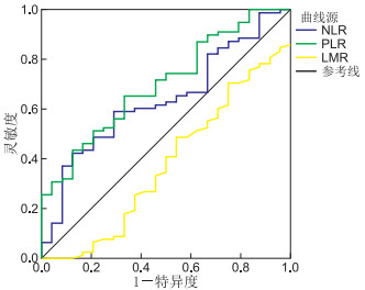
| Citation: | PAN Wan-wan, DONG Meng-hao, YU Fa-zhi, WAN Ling-feng, WU De-lin, LIU Feng, MENG Fan-lun, MA Xiao-peng. Value of peripheral inflammatory markers NLR, PLR and LMR in predicting the efficacy of neoadjuvant chemotherapy for breast cancer[J]. Chinese Journal of General Practice, 2021, 19(9): 1442-1446. doi: 10.16766/j.cnki.issn.1674-4152.002081

|

| [1] |
BRAY F, FERLAY J, SOERJOMATARAM I, et al. Global cancer statistics 2018: GLOBOCAN estimates of incidence and mortality worldwide for 36 cancers in 185 countries[J]. CA Cancer J Clin, 2018, 68(6): 394-424. doi: 10.3322/caac.21492
|
| [2] |
杨辉, 江皓. 新辅助TAC化疗对早期乳腺癌患者疗效、细胞免疫功能和Ki-67的影响[J]. 中华全科医学, 2017, 15(4): 555-557, 671. https://www.cnki.com.cn/Article/CJFDTOTAL-SYQY201704004.htm
|
| [3] |
程阳, 田君, 姚学权, 等. 局部晚期乳腺癌TEC方案新辅助化疗的临床疗效分析[J]. 安徽医学, 2020, 41(9): 1022-1025. doi: 10.3969/j.issn.1000-0399.2020.09.009
|
| [4] |
MOSES C, GARCIA-BLOJ B, HARVEY A R, et al. Hallmarks of cancer: The CRISPR generation[J]. Eur J Cancer, 2018, 93: 10-18. doi: 10.1016/j.ejca.2018.01.002
|
| [5] |
STEFANIUK P, SZYMCZYK A, PODHORECKA M. The neutrophil to lymphocyte and lymphocyte to monocyte ratios as new prognostic factors in hematological malignancies-A narrative review[J]. Cancer Manag Res, 2020, 12: 2961-2977. doi: 10.2147/CMAR.S245928
|
| [6] |
薛国军, 张艳利, 杨光伦. 外周血NLR和PLR预测非luminal型乳腺癌新辅助化疗临床进展的应用[J]. 现代医药卫生, 2019, 35(17): 2629-2632. doi: 10.3969/j.issn.1009-5519.2019.17.011
|
| [7] |
袁茂林, 韩琼, 吴斌. 外周血淋巴细胞和单核细胞比值与乳腺癌新辅助化疗疗效关联性[J]. 中华肿瘤防治杂志, 2020, 27(4): 283-287. https://www.cnki.com.cn/Article/CJFDTOTAL-QLZL202004008.htm
|
| [8] |
BUYUKSIMSEK M, OGUL A, MIRILI C, et al. Inflammatory markers predicting pathological complete response in cases with breast cancer treated by neoadjuvant chemotherapy[J]. Eur J Breast Health, 2020, 16(4): 229-234. doi: 10.5152/ejbh.2020.5556
|
| [9] |
DERKS M G M, VAN DE VELDE C J H. Neoadjuvant chemotherapy in breast cancer: More than just downsizing[J]. Lancet Oncol, 2018, 19(1): 2-3. doi: 10.1016/S1470-2045(17)30914-2
|
| [10] |
BOUGHEY J C, BALLMAN K V, MCCALL L M, et al. Tumor biology and response to chemotherapy impact breast cancer-specific survival in node-positive breast cancer patients treated with neoadjuvant chemotherapy: Long-term follow-up from ACOSOG Z1071(Alliance)[J]. Ann Surg, 2017, 266(4): 667-676. doi: 10.1097/SLA.0000000000002373
|
| [11] |
KLEIN J, TRAN W, WATKINS E, et al. Locally advanced breast cancer treated with neoadjuvant chemotherapy and adjuvant radiotherapy: A retrospective cohort analysis[J]. BMC Cancer, 2019, 19(1): 306. doi: 10.1186/s12885-019-5499-2
|
| [12] |
ALDAWSARI H M, GORAIN B, ALHAKAMY N A, et al. Role of therapeutic agents on repolarisation of tumour-associated macrophage to halt lung cancer progression[J]. J Drug Target, 2020, 28(2): 166-175. doi: 10.1080/1061186X.2019.1648478
|
| [13] |
张美云, 李晓凤, 刘卓. 中性粒细胞与淋巴细胞比值在乳腺癌新辅助化疗中的意义[J]. 癌症进展, 2018, 16(11): 1408-1410. https://www.cnki.com.cn/Article/CJFDTOTAL-AZJZ201811023.htm
|
| [14] |
曲幽, 杜闯, 李靖若. 血小板与淋巴细胞比率对乳腺癌患者新辅助化疗后病理完全缓解的预测价值[J]. 肿瘤基础与临床, 2019, 32(6): 505-510. doi: 10.3969/j.issn.1673-5412.2019.06.012
|
| [15] |
王浩峰, 王耀辉, 陆劲松. 乳腺癌反映炎症状态外周血细胞间比值研究进展[J]. 中华肿瘤防治杂志, 2019, 26(8): 598-602. https://www.cnki.com.cn/Article/CJFDTOTAL-QLZL201908018.htm
|
| [16] |
TAKEUCHI H, KAWANAKA H, FUKUYAMA S, et al. Comparison of the prognostic values of preoperative inflammation-based parameters in patients with breast cancer[J]. PLoS One, 2017, 12(5): e0177137. doi: 10.1371/journal.pone.0177137
|
| [17] |
WARISS B R, DE SOUZA A K, DE AGUIAR S S, et al. Effectiveness of four inflammatory markers in predicting prognosis in 2374 women with breast cancer[J]. Maturitas, 2017, 101: 51-56. doi: 10.1016/j.maturitas.2017.04.015
|
| [18] |
高思铭. 中性粒细胞与淋巴细胞比值预测乳腺癌新辅助化疗疗效及预后的研究[D]. 济南: 山东大学, 2020.
|
| [19] |
OU Q, CHENG J, ZHANG L, et al. The prognostic value of pretreatment neutrophil-to-lymphocyte ratio in breast cancer: Deleterious or advantageous?[J]. Tumour Biol, 2017, 39(6): 1010428317706214. http://www.onacademic.com/detail/journal_1000039945705910_78cc.html
|
| [20] |
彭阳. 外周血中炎症相关细胞比值对乳腺癌患者新辅助化疗疗效的预测作用[D]. 重庆: 重庆医科大学, 2018.
|
| [21] |
郑帅, 毛艳, 贺薇, 等. 乳腺癌患者外周血炎症指标与新辅助化疗疗效的相关性分析[J]. 肿瘤药学, 2020, 10(1): 97-102. https://www.cnki.com.cn/Article/CJFDTOTAL-LIYX202001018.htm
|
| [22] |
周洁, 马景玲, 蒋冬梅, 等. 肿瘤相关炎症指标对乳腺癌新辅助化疗疗效预测价值研究[J]. 中国煤炭工业医学杂志, 2020, 23(6): 626-631. https://www.cnki.com.cn/Article/CJFDTOTAL-ZMGY202006014.htm
|
| [23] |
何洋, 赵伟鹏, 佟仲生. 新辅助化疗对乳腺癌ER、PR、HER-2及Ki-67表达影响的研究进展[J]. 中国肿瘤临床, 2020, 47(22): 1185-1188. https://www.cnki.com.cn/Article/CJFDTOTAL-ZGZL202022017.htm
|
| [24] |
ACS B, ZAMBO V, VIZKELETI L, et al. Ki-67 as a controversial predictive and prognostic marker in breast cancer patients treated with neoadjuvant chemotherapy[J]. Diagn Pathol, 2017, 12(1): 20. doi: 10.1186/s13000-017-0608-5
|
| [25] |
张彦收, 唐甜甜, 周涛, 等. Ki-67表达水平检测在乳腺癌新辅助化疗疗效评估中的价值[J]. 现代肿瘤医学, 2017, 25(14): 2256-2259. https://www.cnki.com.cn/Article/CJFDTOTAL-SXZL201714017.htm
|
Figures(1) / Tables(3)
Address:No. 287, Changhuai Road, Bengbu, Anhui Province, 233004, P.R.ChinaphoneNo:0552-3066635; 0552-3051890Email:zhqkyx@163.com
Copyright © Chinese Journal of General Practice皖ICP备2020018345号-2
Supported by:
Beijing Renhe Information Technology Co., Ltd.


 DownLoad: