| Citation: | ZHA Zheng-wei, GAN Hui-zhong, PENG Qiong, KONG De-run. Diagnostic value of endoscopic ultrasonography for submucosal tumors of digestive tract[J]. Chinese Journal of General Practice, 2022, 20(2): 290-293. doi: 10.16766/j.cnki.issn.1674-4152.002335

|

| [1] |
GRESS F G. Progress in endoscopic ultrasonography[J]. Gastrointest Endosc Clin N Am, 2017, 27(4): 15-16.
|
| [2] |
周维霞, 丁科枫, 殷国建, 等. 超声内镜对结直肠黏膜下病变的诊断价值[J]. 中国内镜杂志, 2017, 23(6): 92-97. https://www.cnki.com.cn/Article/CJFDTOTAL-ZGNJ201706019.htm
|
| [3] |
施丹, 李文, 石磊, 等. 内镜超声在结直肠黏膜下隆起性病变的应用分析[J]. 中华消化内镜杂志, 2020, 37(6): 415-419. doi: 10.3760/cma.j.cn321463-20191114-00766
|
| [4] |
AlPERT L, Al-SABTI R, GRAHAM R P, et al. Smooth muscle tumors of the gastrointestinal tract: An analysis of prognostic features in 407 cases[J]. Mod Pathol, 2020, 33: 1410-1419. doi: 10.1038/s41379-020-0492-5
|
| [5] |
丁艳乐, 丰义宽, 周新玲, 等. 超声内镜在上消化道黏膜下肿瘤诊断价值[J]. 胃肠病学和肝病学杂志, 2017, 26(10): 1146-1149. doi: 10.3969/j.issn.1006-5709.2017.10.020
|
| [6] |
刘铁权. 超声内镜在上消化黏膜下肿瘤诊治中的应用分析[J]. 航空航天医学杂志, 2020, 31(11): 1335-1336. doi: 10.3969/j.issn.2095-1434.2020.11.027
|
| [7] |
吴巍, 范嵘, 谭继宏, 等. 内镜超声对消化道黏膜下肿瘤内镜术前评估的价值和局限性[J]. 中华消化内镜杂志, 2019, 36(7): 491-494. doi: 10.3760/cma.j.issn.1007-5232.2019.07.007
|
| [8] |
OKANOUE S, IWAMURO M, TANAKA T, et al. Scoring systems for differentiating gastrointestinal stromal tumors and schwannomas from leiomyomas in the stomach[J]. Medicine (Baltimore), 2021, 100(40): 1-6. http://oldmed.wanfangdata.com.cn/Paper/Detail/PeriodicalPaper_PM34622886
|
| [9] |
KAMATA K, TAKENAKA M, KITANO M, et al. Contrast-enhanced harmonic endoscopic ultrasonography for differential diagnosis of submucosal tumors of the upper gastrointestinal tract[J]. J Gastroenterol Hepatol, 2017, 32(10): 1686-1692. doi: 10.1111/jgh.13766
|
| [10] |
赵丽莎, 龙辉, 郝顺心. 超声胃镜对上消化道隆起性病变的诊疗价值[J]. 临床内科杂志, 2017, 34(9): 606-607. doi: 10.3969/j.issn.1001-9057.2017.09.008
|
| [11] |
SU Q, PENG J, CHEN X, et al. Role of endoscopic ultrasonography for differential diagnosis of upper gastrointestinal submucosal lesions[J]. BMC Gastroenterol, 2021, 21(1): 365. doi: 10.1186/s12876-021-01945-9
|
| [12] |
杨智炜, 王胜炳, 汪福群, 等. 108例食管、胃黏膜下隆起超声内镜诊断分析[J]. 中国医药科学, 2017, 7(5): 108-110. doi: 10.3969/j.issn.2095-0616.2017.05.031
|
| [13] |
施丹, 李文, 石磊, 等. 内镜超声在结直肠黏膜下隆起性病变的应用分析[J]. 中华消化内镜杂志, 2020, 37(6): 415-419. doi: 10.3760/cma.j.cn321463-20191114-00766
|
| [14] |
王玮, 罗丹, 李启祥, 等. 胃间质瘤不同危险等级的评估因素及超声内镜的应用价值[J]. 现代消化及介入诊疗, 2019, 24(3): 303-306. doi: 10.3969/j.issn.1672-2159.2019.03.024
|
| [15] |
黄军, 郑海伦, 王启之, 等. 微探头超声内镜辅助内镜治疗消化道黏膜下肿物291例临床分析[J]. 中华全科医学, 2017, 15(12): 2038-2041. https://www.cnki.com.cn/Article/CJFDTOTAL-SYQY201712012.htm
|
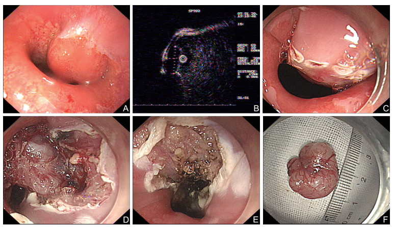
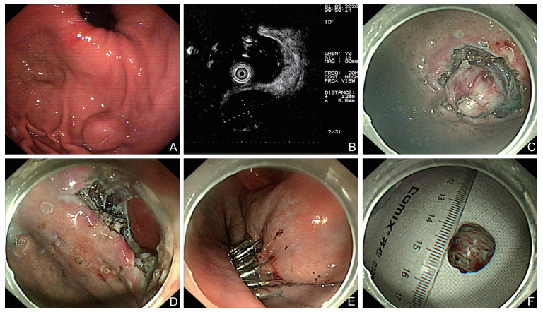
Figures(2) / Tables(1)
Address:No. 287, Changhuai Road, Bengbu, Anhui Province, 233004, P.R.ChinaphoneNo:0552-3066635; 0552-3051890Email:zhqkyx@163.com
Copyright © Chinese Journal of General Practice皖ICP备2020018345号-2
Supported by:
Beijing Renhe Information Technology Co., Ltd.


 DownLoad: