

Value of strain elastography and shear wave elastography combined with BI-RADS in determining the nature of breast masses
-
摘要:
目的 探讨应变式弹性成像和剪切波弹性成像技术分别联合乳腺超声影像报告和数据系统(BI-RADS)对乳腺肿块性质良恶性的诊断价值。 方法 回顾性分析2021年4月—2023年3月合肥市第二人民医院收治的105例乳腺肿块患者(共105个病灶)的临床病理资料,术前接受常规超声检查、应变式弹性成像和剪切波弹性成像检查。以病理结果为金标准将病灶分为良性组(64例)和恶性组(41例),比较2组弹性应变率比值(SR)、剪切波最大弹性模量值(Emax),绘制ROC曲线评估BI-RADS、SR、Emax及联合诊断恶性乳腺肿块的效能。 结果 105例乳腺肿块患者经病理检查显示,共41例(39.05%)恶性肿块,64例(60.95%)良性肿块。BI-RADS分类结果显示,2类10例,3类16例,4a类21例,4b类19例,4c类18例,5类21例。以4b分类及以上诊断为恶性,BI-RADS分类诊断恶性乳腺肿块的灵敏度为92.68%,特异度为68.75%。恶性组SR、Emax值均高于良性组(P<0.05)。ROC曲线分析显示,SR、Emax诊断恶性乳腺肿块的AUC分别为0.739、0.784,最佳截断值分别为4.17%、98.21 kPa;SR+Emax联合诊断的AUC为0.861,灵敏度为70.73%,特异度为92.19%,约登指数为0.629,联合诊断效能高于单项诊断(P<0.05)。ROC曲线分析显示,SR+BI-RADS、Emax+BI-RADS、SR+Emax+BI-RADS诊断恶性乳腺肿块的AUC分别为0.826、0.830、0.892,SR+Emax+BI-RADS诊断效能高于SR+BI-RADS、Emax+BI-RADS、BI-RADS(P<0.05)。 结论 SR+Emax+BI-RADS联合诊断能明显提高乳腺癌的诊断效能,提高了BI-RADS分类诊断的特异度。 -
关键词:
- 应变式弹性成像 /
- 剪切波弹性成像 /
- 乳腺超声影像报告和数据系统 /
- 乳腺肿块 /
- 诊断
Abstract:Objective To investigate the diagnostic value of strain elastic imaging and shear wave elastic imaging combined with breast imaging reporting and data system (BI-RADS) in benign and malignant breast masses. Methods The clinicopathological data of 105 patients with breast masses (105 lesions in total) admitted to our hospital from April 2021 to March 2023 were reviewed. Preoperatively, they underwent routine ultrasonography, strain elastography, and shear wave elastography. The lesions were classified into benign and malignant groups using the pathological findings as the gold standard. Elastic strain ratio (SR), and shear wave maximum elastic modulus value (Emax) were compared between the two groups. The efficacy of BI-RADS, SR, Emax, and the combined diagnosis of malignancy of breast masses was evaluated by plotting the working characteristic ROC curves of the subjects. Results A total of 105 breast masses were examined pathologically showing 41 (39.05%) malignant and 64 (60.95%) benign masses. BI-RADS classification showed 10 cases in category 2, 16 cases in category 3, 21 cases in category 4a, 19 cases in category 4b, 18 cases in category 4c, and 21 cases in category 5. With a diagnosis of malignancy at 4b classification and above, the sensitivity of the BI-RADS classification for diagnosing malignant breast masses was 92.68% and the specificity was 68.75%. The SR and Emax values were higher in the malignant group than in the benign group (P < 0.05). ROC curve analysis showed that the AUCs of SR and Emax for diagnosing malignant breast masses were 0.739 and 0.784, and the optimal cut-off values were 4.17% and 98.21 kPa, respectively, and the AUCs of SR+Emax combined diagnosis were 0.861, with a sensitivity of 70.73% and a specificity of 92.19%, and a Yoden index of 0.629, which showed that the efficacy of the combination was higher than that of the single diagnosis (P < 0.05). ROC curve analysis showed that the AUCs of SR+BI-RADS, Emax+BI-RADS, and SR+Emax+BI-RADS for diagnosing malignant breast masses were 0.826, 0.830, and 0.892, respectively, and the diagnostic efficacy of SR+Emax+BI-RADS was higher than that of SR+BI-RADS, Emax+BI-RADS, and BI-RADS (P < 0.05). Conclusion The combined diagnosis of SR+Emax+BI-RADS can significantly improve the diagnostic efficacy of breast cancer, and improve the specificity of BI-RADS classification diagnosis. -

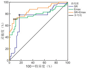
图 1 SR、Emax及联合诊断恶性乳腺肿块的ROC曲线
Figure 1. ROC curves of SR, Emax, and their combined diagnosis for malignant breast masses

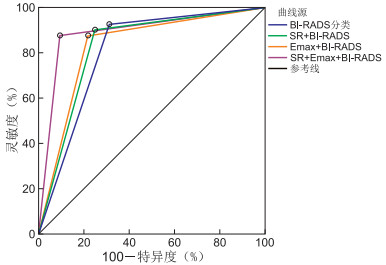
图 2 SR、Emax联合BI-RAD诊断恶性乳腺肿块的ROC曲线
Figure 2. ROC curve of SR, Emax combined with BI-RAD in diagnosing malignant breast masses

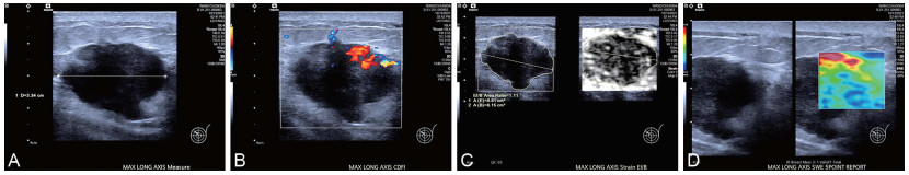
图 3 浸润性导管癌超声检查图像
注:患者49岁,浸润性导管癌。A为二维超声图,最长径为3.34 cm,形态不规则,边界不清,内部见点状强回声;B为彩色多普勒超声显示肿块内部丰富血流信号;C为应变式弹性成像图,SR值为5.59%;D为剪切波弹性成像图,病灶显示红色蓝色相间,Emax值为101.2 kPa。
Figure 3. Ultrasonographic image of invasive ductal carcinoma

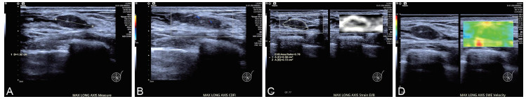
图 4 纤维腺瘤样结节超声检查图像
注:患者42岁,纤维腺瘤样结节。A为二维超声图,最长径为1.52 cm;B为彩色多普勒超声显示肿块内点状血流信号;C为应变式弹性成像图,SR值为1.27%;D为剪切波弹性成像图,病灶显示较为均匀的绿色,Emax值为62.4 kPa。
Figure 4. Ultrasonographic images of fibroadenomatous nodule
表 1 BI-RADS分类与病理诊断对乳腺肿块性质的诊断结果比较(例)
Table 1. Comparison of BI-RADS classification and pathological diagnosis for the features of breast lumps (cases)
BI-RADS分类 病理结果 合计 恶性 良性 恶性 38 20 58 良性 3 44 47 合计 41 64 105  下载: 导出CSV
表 2 良恶性组乳腺肿块患者SR、Emax值比较(x±s)
Table 2. Comparison of SR and Emax values between patients with benign or malignant breast masses (x±s)
组别 例数 SR(%) Emax(kPa) 良性组 64 3.31±0.97 86.36±10.25 恶性组 41 4.06±0.91 103.39±20.05 t值 3.384 5.390 P值 0.001 <0.001
下载: 导出CSV
表 3 SR、Emax值对乳腺肿块良恶性的诊断价值
Table 3. Diagnostic value of SR and Emax values for benign and malignant breast masses
项目 AUC 95% CI 最佳截断值 灵敏度(%) 特异度(%) 约登指数 P值 SR 0.739 0.644~0.820 4.17% 78.05(32/41) 82.81(53/64) 0.609 <0.001 Emax 0.784 0.693~0.858 98.21 kPa 70.73(29/41) 89.06(57/64) 0.598 <0.001 SR+Emax 0.861 0.780~0.921 70.73(29/41) 92.19(59/64) 0.629 <0.001
下载: 导出CSV
表 4 SR、Emax联合BI-RADS对乳腺肿块良恶性的诊断价值
Table 4. Diagnostic value of SR, Emax combined with BI-RADS for benign and malignant breast masses
项目 AUC 95% CI 灵敏度(%) 特异度(%) 约登指数 P值 BI-RADS 0.807 0.719~0.878 92.68(38/41) 68.75(44/64) 0.614 <0.001 SR+BI-RADS 0.826 0.740~0.893 90.24(37/41) 75.00(48/64) 0.652 <0.001 Emax+BI-RADS 0.830 0.744~0.896 87.80(36/41) 78.12(50/64) 0.659 <0.001 SR+Emax+BI-RADS 0.892 0.817~0.944 87.80(36/41) 90.62(58/64) 0.784 <0.001
下载: 导出CSV
-
[1] KATSURA C, OGUNMWONYI I, KANKAM H K, et al. Breast cancer: presentation, investigation and management[J]. Br J Hosp Med(Lond), 2022, 83(2): 1-7. [2] 郭赛灵, 朱爽爽, 邢伟, 等. 基于2013版BI-RADS术语及关于CEM术语的补充规定探讨CEM与MRI鉴别乳腺良性与恶性病变的价值[J]. 中华放射学杂志, 2023, 57(7): 762-770.GUO S L, ZHU S S, XING W, et al. Diagnostic value of CEM and MRI in differentiating benign and malignant breast lesions based on the 2013 BI-RADS lexicon and the supplement on CEM[J]. Chinese Journal of Radiology, 2023, 57(7): 762-770. [3] KAMAL A M, SAKORIKAR T, PAL U M, et al. Engineering approaches for breast cancer diagnosis: a review[J]. IEEE Rev Biomed Eng, 2023, 16: 687-705. doi: 10.1109/RBME.2022.3181700 [4] PFOB A, SIDEY-GIBBONS C, BARR R G, et al. Intelligent multi-modal shear wave elastography to reduce unnecessary biopsies in breast cancer diagnosis (INSPiRED 002): a retrospective, international, multicentre analysis[J]. Eur J Cancer, 2022, 177: 1-14. doi: 10.1016/j.ejca.2022.09.018 [5] 冯淑, 李阳, 靳鹏, 等. 超声萤火虫联合超微血管成像技术对比X线钼靶诊断乳腺疾病的价值[J]. 中华全科医学, 2021, 19(12): 2098-2101. doi: 10.16766/j.cnki.issn.1674-4152.002246FENG S, LI Y, JIN P, et al. Value of micropure combined with superb microvascular imaging compared with X-ray molybdenum target for the diagnosis of breast diseases[J]. Chinese Journal of General Practice, 2021, 19(12): 2098-2101. doi: 10.16766/j.cnki.issn.1674-4152.002246 [6] HUANG Q, YE L. Multi-task/single-task joint learning of ultrasound BI-RADS features[J]. IEEE Trans Ultrason Ferroelectr Freq Control, 2022, 69(2): 691-701. doi: 10.1109/TUFFC.2021.3132933 [7] EGHTEDARI M, CHONG A, RAKOW-PENNER R, et al. Current status and future of BI-RADS in multimodality imaging, from the AJR special series on radiology reporting and data systems[J]. AJR Am J Roentgenol, 2021, 216(4): 860-873. doi: 10.2214/AJR.20.24894 [8] SARHANGI N, HAJJARI S, HEYDARI S F, et al. Breast cancer in the era of precision medicine[J]. Mol Biol Rep, 2022, 49(10): 10023-10037. doi: 10.1007/s11033-022-07571-2 [9] GOLATTA M, PFOB A, BVSCH C, et al. The potential of shear wave elastography to reduce unnecessary biopsies in breast cancer diagnosis: an international, diagnostic, multicenter trial[J]. Ultraschall Med, 2023, 44(2): 162-168. doi: 10.1055/a-1543-6156 [10] SACCENTI L, MELLON C M, SCHOLER M, et al. Combining b2500 diffusion-weighted imaging with BI-RADS improves the specificity of breast MRI[J]. Diagn Interv Imaging, 2023, 104(9): 410-418. doi: 10.1016/j.diii.2023.05.001 [11] FANG Y, ZHOU Y. Diagnosis of breast cancer lesion using ultrasound images, elastography, and Ki-67 protein cell proliferation index[J]. Cell Mol Biol(Noisy-le-grand), 2023, 69(4): 16-23. doi: 10.14715/cmb/2023.69.4.3 [12] XU Y J, GONG H L, HU B, et al. Role of "Stiff Rim" sign obtained by shear wave elastography in diagnosis and guiding therapy of breast cancer[J]. Int J Med Sci, 2021, 18(15): 3615-3623. doi: 10.7150/ijms.64243 [13] KOKUBU Y, YAMADA K, TANABE M, et al. Evaluating the usefulness of breast strain elastography for intraductal lesions[J]. J Med Ultrason(2001), 2021, 48(1): 63-70. doi: 10.1007/s10396-020-01070-2 [14] PEKER A, BALCI P, BASARA AKIN I, et al. Shear-wave elastography-guided core needle biopsy for the determination of breast cancer molecular subtype[J]. J Ultrasound Med, 2021, 40(6): 1183-1192. doi: 10.1002/jum.15499 [15] 沈朝明, 陈淑霞, 邬午龙, 等. 剪切波弹性成像对乳腺癌良恶性的鉴别诊断及误诊分析[J]. 中华全科医学, 2020, 18(4): 634-637. doi: 10.16766/j.cnki.issn.1674-4152.001317SHEN Z M, CHEN S X, WU W L, et al. Differential diagnosis and misdiagnosis of benign and malignant breast cancer by shear wave elastography[J]. Chinese Journal of General Practice, 2020, 18(4): 634-637. doi: 10.16766/j.cnki.issn.1674-4152.001317 [16] 唐燕, 谷颖. 超声筛查乳腺恶性非肿块型病变的研究现状[J]. 中华妇幼临床医学杂志(电子版), 2022, 18(4): 379-386. https://www.cnki.com.cn/Article/CJFDTOTAL-ZHFY202204002.htmTANG Y, GU Y. Current research status of breast malignant non-mass lesion by ultrasound screening[J]. Chinese Journal of Obstetrics & Gynecology and Pediatrics(Electronic Edition), 2022, 18(4): 379-386. https://www.cnki.com.cn/Article/CJFDTOTAL-ZHFY202204002.htm [17] LEE E J, CHANG Y W. Prospective analysis of breast masses using the combined score for quantitative ultrasonography parameters[J]. Sci Rep, 2022, 12(1): 16205. DOI: 10.1038/s41598-022-19971-2. [18] LI J, SUN B, LI Y, et al. Correlation analysis between shear-wave elastography and pathological profiles in breast cancer[J]. Breast Cancer Res Treat, 2023, 197(2): 269-276. -

 点击查看大图
计量
- 文章访问数: 829
- HTML全文浏览量: 610
- PDF下载量: 8
- 被引次数: 0
