

Clinical study of Modified Taohong Siwu Decoction in the treatment of diabetic macular edema of Qi-Yin deficiency combined with blood stasis
-
摘要:
目的 观察加味桃红四物汤(MTSD)联合雷珠单抗治疗糖尿病黄斑水肿(DME)的疗效及对视网膜微循环的影响。 方法 选取2022年9月—2023年9月上海中医药大学附属曙光医院眼科收治的DME患者100例(100眼),按随机数字表法分为2组,各50例50眼,对照组予玻璃体腔注射雷珠单抗,治疗组在对照组基础上口服MTSD,随访3个月。观察2组治疗前后的中医证候积分、最佳矫正视力(BCVA)、黄斑中心凹厚度(CMT)、应用光学相干断层扫描血管成像测量三分区四方位的视网膜浅层血管长度密度(VD)、浅层血管灌注密度(PD)。 结果 与对照组比较,治疗组中医证候疗效更好(P < 0.05)。治疗组治疗后的BCVA(4.80±0.21)大于对照组(4.64±0.30, F=34.857, P < 0.05)。治疗后2组CMT均降低(P < 0.01),但组间差异无统计学意义(P>0.05)。治疗组治疗后内层区上方和下方以及外层区上方和下方的VD及PD均较治疗前增加(P < 0.05),并且除颞侧外,治疗组中心区、内层区和外层区的上方、鼻侧、下方VD及PD均较对照组增加(P < 0.05)。 结论 与单纯玻璃体腔注射雷珠单抗相比,联合MTSD治疗气阴两虚兼血瘀阻络型DME可以改善患者的不适症状,提高视力,改善除了颞侧以外的视网膜微循环,是辅助治疗DME的有效方法。 -
关键词:
- 糖尿病黄斑水肿 /
- 加味桃红四物汤 /
- 雷珠单抗 /
- 视网膜微循环 /
- 光学相干断层扫描血管成像
Abstract:Objective To observe the clinical efficacy of Modified Taohong Siwu Decoction (MTSD) combined with Ranibizumab in the treatment of diabetic macular edema (DME) and its influence on retinal microcirculation. Methods A total of 100 patients (100 eyes) with DME in the Ophthalmology Department of Shuguang Hospital from September 2022 to September 2023 were picked and randomly divided into two groups according to the random list method, with 50 cases and 50 eyes in each group. The control group was treated with intravitreal injection of Ranibizumab. The treatment group was treated with oral MTSD combined with intravitreal injection of Ranibizumab, and followed up for 3 months. TCM syndrome score, best corrected visual acuity, central macular thickness, superficial vascular density and superficial perfusion density in three zones and four places by OCTA in the two groups were measured and compared before and after treatment. Results The improvement in TCM symptoms in the treatment group was better than that in the control group (P < 0.05). The BCVA of the treatment group after treatment (4.80±0.21) was greater than that of the control group (4.64±0.30, F=34.857, P < 0.05). After treatment, both groups could reduce CMT (P < 0.001), but there was no statistically significant difference in the groups (P>0.05). After treatment in the treatment group, the VD and PD above and below the inner layer area and above and below the outer layer area increased compared with those before treatment (all P < 0.05). And except for the temporal side, the VD and PD in the central area, the upper part of the inner and outer layers, the nasal side and the lower part of the treatment group increased compared with the control group (P < 0.05). Conclusion Compared with simple intravitreal injection of Ranibizumab, MTSD combined in the treatment of DME can improve the discomfort symptoms of patients with Qi-Yin deficiency combined with blood stasis, improve the visual acuity, and improve the retinal microcirculation except the temporal side, which is an effective method for the adjuvant treatment of DME. -

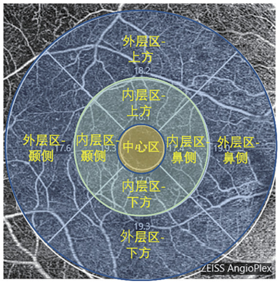
图 1 以右眼为例的OCTA观察视网膜浅层血管量化指标的分区指示图
注: 蓝色为外层区, 绿色为内层区, 橙色为中心区。
Figure 1. Zonal indication map of quantitative superficial retinal vessel parameters obtained by OCTA, using the right eye as an example
表 1 2组DME患者中医证候疗效比较
Table 1. Comparison of TCM syndrome improvement between the two groups of DME patients
组别 例数 痊愈
(例)显效
(例)有效
(例)无效
(例)总有效率
(%)对照组 50 0 4 30 16 68.00 治疗组 50 0 11 29 10 80.00 注:2组中医证候疗效比较,Z=-2.014,P=0.044。  下载: 导出CSV
表 2 2组DME患者BCVA比较(x±s)
Table 2. Comparison of BCVA between the two groups of DME patients (x±s)
组别 例数 治疗前 治疗后 t值 P值 对照组 50 4.56±0.32 4.64±0.30 4.802 < 0.001 治疗组 50 4.57±0.29 4.80±0.21 8.363 < 0.001 统计量 0.193a 34.857b P值 0.847 < 0.001 注:a为t值,b为F值。
下载: 导出CSV
表 3 2组DME患者治疗前后CMT比较(x±s,μm)
Table 3. Comparison of CMT between the two groups of DME patients (x±s, μm)
组别 例数 治疗前 治疗后 t值 P值 对照组 50 323.24±117.48 270.06±49.11 3.027 0.004 治疗组 50 349.10±144.99 279.98±59.79 3.946 < 0.001 统计量 0.980a 0.440b P值 0.330 0.509 注:a为t值,b为F值。
下载: 导出CSV
表 4 2组DME患者VD各分区比较(%)
Table 4. Comparison of VD in each zone between the two groups of DME patients (%)
组别 例数 时间 中心区[M(P25, P75)] 内层区(x±s) 外层区(x±s) VD上方 VD鼻侧 VD下方 VD颞侧 VD上方 VD鼻侧 VD下方 VD颞侧 对照组 50 治疗前 4.60(1.77, 8.05) 11.00±5.07 11.40±5.57 11.23±5.39 10.73±5.32 12.29±4.53 14.09±5.14 11.68±4.84 10.26±5.03 治疗后 3.65(1.40, 6.15) 10.27±4.76 10.15±4.97 9.88±4.73 10.15±5.10 12.22±3.83 14.36±4.15 11.50±4.19 10.33±4.75 治疗组 50 治疗前 4.85(1.70, 8.62) 11.41±5.62 11.70±5.64 10.90±5.73 10.43±6.22 12.46±4.68 15.57±4.17 12.22±4.85 10.41±5.75 治疗后 5.25(2.80, 8.80)a 13.06±4.48ab 13.15±4.89a 12.90±4.22ab 12.04±5.28 14.25±3.82ab 16.56±3.83a 13.97±3.91ab 11.66±4.95 注:与对照组比较,aP < 0.05;与治疗前比较,bP < 0.05。
下载: 导出CSV
表 5 2组DME患者PD各分区比较(%)
Table 5. Comparison of PD in each zone between the two groups of DME patients (%)
组别 例数 时间 中心区
[M(P25, P75)]内层区(x±s) 外层区(x±s) PD上方 PD鼻侧 PD下方 PD颞侧 PD上方 PD鼻侧 PD下方 PD颞侧 对照组 50 治疗前 0.090(0.031, 0.168) 0.257±0.125 0.260±0.137 0.260±0.137 0.243±0.133 0.299±0.122 0.344±0.131 0.279±0.136 0.240±0.131 治疗后 0.074(0.032, 0.128) 0.237±0.118 0.235±0.123 0.228±0.115 0.235±0.126 0.295±0.100 0.345±0.108 0.277±0.111 0.247±0.120 治疗组 50 治疗前 0.104(0.034, 0.179) 0.265±0.131 0.282±0.140 0.282±0.140 0.243±0.150 0.308±0.122 0.375±0.115 0.293±0.126 0.252±0.146 治疗后 0.121(0.057, 0.193)a 0.310±0.121ab 0.306±0.130b 0.304±0.111ab 0.281±0.132 0.346±0.108ab 0.401±0.106a 0.345±0.110ab 0.281±0.131 注:与对照组比较,aP < 0.05;与治疗前比较,bP < 0.05。
下载: 导出CSV
-
[1] 吴卫明, 包蕾, 彭志佳, 等. 糖尿病视网膜病变病人血清STAT1和NQO1水平与视力损伤程度的关系[J]. 青岛大学学报(医学版), 2025, 61(1): 69-72.WU W M, BAO L, PENG Z J, et al. Correlation of the levels of serum activator of transcription factor 1 and quinone oxidoreductase 1 with the severity of visual impairment in patients with diabetic retinopathy[J]. Journal of Qingdao University(medical sciences), 2025, 61(1): 69-72. [2] 夏静, 陈佳玉, 杨爱萍, 等. 25G+PPV联合术前玻璃体腔注射康柏西普治疗增生型糖尿病视网膜病变[J]. 国际眼科杂志, 2023, 23(2): 294-298.XIA J, CHEN J Y, YANG A P, et al. Effect of 25G+pars plana vitrectomy combined with preoperative intravitreal injection of Conbercept on proliferative diabetic retinopathy[J]. International Eye Science, 2023, 23(2): 294-298. [3] 殷小敏, 薛寒, 翟丽萍. 康柏西普对合并黄斑水肿的糖尿病视网膜病变患者的影响[J]. 中国医药导报, 2022, 19(33): 100-103.YIN X M, XUE H, ZHAI L P. Influence of Conbercept on diabetic retinopathy patients with macular edema[J]. China Medical Herald, 2022, 19(33): 100-103. [4] 黄栊瑢, 付美林, 刘志敏, 等. 从AQP4/Kir4.1的表达和分布探讨水平衡在糖尿病视网膜水肿中的机制与研究进展[J]. 中国中医眼科杂志, 2023, 33(10): 973-977.HUANG L R, FU M L, LIU Z M, et al. Mechanisms and research progress of fluid balance in diabetic macular edema: Distribution of AQP4 and Kir4.1[J]. China Journal of Chinese Ophthalmology, 2023, 33(10): 973-977. [5] 钟晴雅, 何恒倩, 张军涛, 等. 糖尿病性黄斑水肿的治疗新进展[J]. 现代实用医学, 2023, 35(8): 1109-1112.ZHONG Q Y, HE H Q, ZHANG J T, et al. New advances in the treatment of diabetic macular edema[J]. Modern Practical Medicine, 2023, 35(8): 1109-1112. [6] HATAMNEJAD A, ORR S, DADAK R, et al. Anti-VEGF and steroid combination therapy relative to anti-VEGF mono therapy for the treatment of refractory DME: a systematic review of efficacy and meta-analysis of safety[J]. Acta Ophthalmol, 2024, 102(3): e204-e214. DOI: 10.1111/aos.15724. [7] 董泽英, 杨青青, 潘艳, 等. IVR+PRP联合PPV治疗PDR对患者眼表功能、屈光状态及肾功能的影响[J]. 中华全科医学, 2024, 22(4): 592-596. doi: 10.16766/j.cnki.issn.1674-4152.003458DONG Z Y, YANG Q Q, PAN Y, et al. Clinical effect of IVR+PRP combined with PPV on ocular surface function, refractive status and renal function in patients with proliferative diabetic retinopathy[J]. Chinese Journal of General Practice, 2024, 22(4): 592-596. doi: 10.16766/j.cnki.issn.1674-4152.003458 [8] GURUNG R L, FITZGERALD L M, LIU E, et al. Predictive factors for treatment outcomes with intravitreal anti-vascular endothelial growth factor injections in diabetic macular edema in clinical practice[J]. Int J Retina Vitreous, 2023, 9(1): 23. DOI: 10.1186/s40942-023-00453-0. [9] ULUDAG G, HASSAN M, MATSUMIVA W, et al. Efficacy and safety of intravitreal anti-VEGF therapy in diabetic retinopathy: what we have learned and what should we learn further?[J]. Expert Opin Biol Ther, 2022, 22(10): 1275-1291. doi: 10.1080/14712598.2022.2100694 [10] 杨宇琴, 刘吉民, 李景景, 等. 加味桃红四物汤联合激光治疗重度非增生型糖尿病视网膜病变的临床研究[J]. 上海中医药杂志, 2023, 57(10): 54-58.YANG Y Q, LIU J M, LI J J, et al. Clinical study on treatment of severe non-proliferative diabetic retinopathy with modified Taohong Siwu Decoction and laser therapy[J]. Shanghai Journal of Traditional Chinese Medicine, 2023, 57(10): 54-58. [11] 范先群, 颜华. 眼科学[M]. 10版. 北京: 人民卫生出版社, 2024: 159-160.FAN X Q, YAN H. Ophthalmology[M]. The 10th Edition. Beijing: People' s Medical Publishing House, 2024: 159-160. [12] EHLERS J P, UCHIDA A, SEVGI D D, et al. Retinal fluid volatility associated with interval tolerance and visual outcomes in diabetic macular edema in the VISTA Phase Ⅲ Trial[J]. Am J Ophthalmol, 2021, 224(4): 217-227. [13] 张梅珍, 刘求红. 糖尿病无明显视力下降患者黄斑区血流密度观察及中医证型研究[J]. 新中医, 2023, 55(18): 54-60.ZHANG M Z, LIU Q H. Observation on macular blood flow density of patients with no obvious sight loss caused by diabetes and study on traditional chinese medicine syndrome types[J]. New Chinese Medicine, 2023, 55(18): 54-60. [14] 宋海姣, 金明. 眼底黄斑水肿之三阴辨治思考[J]. 中国医药导报, 2022, 19(3): 119-122.SONG H J, JIN M. Thoughts on differentiation and treatment of three yin-meridian in fundus macular edema[J]. China Medical Herald, 2022, 19(3): 119-122. [15] 李晓东, 高彦, 王艳青, 等. 不同程度糖尿病视网膜病变患者黄斑区血流的变化及意义: 基于OCTA的评价[J]. 眼科新进展, 2021, 41(4): 338-342.LI X D, GAO Y, WANG Y Q, et al. Changes and significance of macular blood flow in patients with diabetic retinopathy of different severity: based on optical coherence tomography angiography[J]. Rec Adv Ophthalmol, 2021, 41(4): 338-342. [16] 许厚银, 郎胜坤. OCTA对糖尿病视网膜病变患者视网膜微血管检测[J]. 国际眼科杂志, 2022, 22(2): 327-330.XU H Y, LANG S K. Detection of retinal microvessels in patients with diabetic retinopathy by OCTA[J]. International Eye Science, 2022, 22(2): 327-330. [17] 李海东, 方伟, 吴素兰, 等. 非增生期糖尿病视网膜病变患者黄斑区血流密度变化: 基于OCTA的定量分析[J]. 眼科新进展, 2021, 41(2): 170-173.LI H D, FANG W, WU S L, et al. Quantitative analysis of macular vessel density changes in non-proliferative diabetic retinopathy through optical coherence tomography angiography[J]. Recent Advances in Ophthalmology, 2021, 41(2): 170-173. [18] 霍倩倩, 张金燕, 杨凯莉, 等. 不同分层、不同区域增生型糖尿病视网膜病变患眼视网膜毛细血管无灌注区的分布特征: 基于全域SS-OCTA的研究[J]. 眼科新进展, 2023, 43(6): 472-475.HUO Q Q, ZHANG J Y, YANG K L, et al. Distribution characteristics of the retinal capillary non-perfusion area at different layers and zones in patients with proliferative diabetic retinopathy based on full-range swept-source optical coherence tomography angiography[J]. Recent Advances in Ophthalmology, 2023, 43(6): 472-475. -

 点击查看大图
计量
- 文章访问数: 4
- HTML全文浏览量: 2
- PDF下载量: 0
- 被引次数: 0
