



Study on the expression characteristics, prognostic value, and potential mechanisms of TMEM63C in lung adenocarcinoma
-
摘要:
目的 分析跨膜蛋白63C(TMEM63C)在肺腺癌(LUAD)组织中的表达及其与患者预后的关系,并探讨其对LUAD细胞增殖、迁移和侵袭的影响及作用机制。 方法 采用RT-qPCR和Western Blotting检测TMEM63C在LUAD细胞中的表达。采用GEPIA、MetaIntegrator、Kaplan-Meier plotter数据库和Cox回归分析研究TMEM63C在LUAD组织中的表达及预后价值。采用TIMER、LinkedOmics和Metasacape数据库进行免疫浸润和富集分析。siRNA干扰后,采用CCK-8、克隆形成、划痕、Transwell和Western Blotting实验评估细胞增殖、迁移、侵袭及PI3K/AKT蛋白表达变化。 结果 TMEM63C在LUAD中高表达且高表达患者总生存率更高(P<0.05)。多因素Cox回归分析结果显示,TMEM63C高表达是LUAD患者总生存期的独立保护因素[HR(95% CI) : 0.798(0.686~0.928), P=0.003]。TMEM63C表达与NK细胞(r=0.275)和树突状细胞(r=0.188)浸润呈正相关关系,与中性粒细胞(r=-0.106)浸润呈负相关关系(P<0.05)。下调TMEM63C表达促进LUAD细胞增殖、迁移、侵袭并增加PI3K/AKT蛋白表达(P<0.05)。 结论 TMEM63C在LUAD中高表达且与患者良好预后相关,机制可能与调节肿瘤微环境及抑制PI3K/AKT信号通路有关。 Abstract:Objective To analyze the expression of transmembrane protein 63C (TMEM63C) in lung adenocarcinoma (LUAD), its relationship with patient prognosis, and its effects on the proliferation, migration, and invasion of LUAD cells, as well as the underlying molecular mechanisms. Methods TMEM63C expression in LUAD cells was detected by RT qPCR and Western Blotting. Its expression and prognostic value in LUAD tissues were analyzed using the GEPIA, MetaIntegrator, Kaplan Meier plotter databases, and Cox regression. Immune infiltration and enrichment analyses were performed with the TIMER, LinkedOmics, and Metascape databases. Following siRNA interference, CCK-8, colony formation, wound healing, Transwell, and Western Blotting assays were used to assess changes in cell proliferation, migration, invasion, and PI3K/AKT protein expression. Results TMEM63C was highly expressed in LUAD, and elevated expression was significantly associated with improved overall survival (P < 0.05). Multivariate Cox regression analysis showed that high expression of TMEM63C was an independent protective factor for overall survival in LUAD patients [HR (95% CI): 0.798 (0.686~0.928), P=0.003]. TMEM63C expression was positively correlated with infiltration of NK cells (r=0.275) and dendritic cells (r=0.188), and negatively correlated with neutrophil infiltration (r=-0.106, P < 0.05). Knockdown of TMEM63C significantly promoted LUAD cell proliferation, migration, and invasion, accompanied by increased expression of PI3K and AKT proteins (P < 0.05). Conclusion TMEM63C is highly expressed in LUAD and is associated with a favorable patient prognosis. Its tumor-suppressive effects may be mediated through modulating of the tumor microenvironment and inhibiting of the PI3K/AKT signaling pathway. -

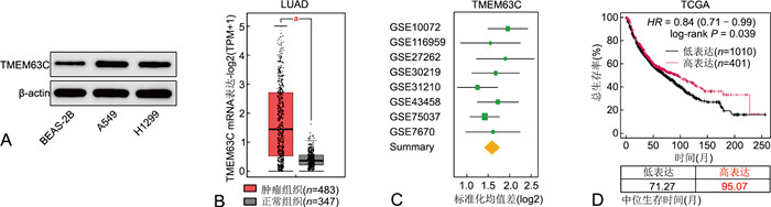
图 1 LUAD中TMEM63C mRNA和蛋白质表达及预后分析
注:A为Western blotting分析TMEM63C在人正常支气管上皮细胞BEAS-2B和LUAD细胞系A549和H1299中的表达;B为GEPIA数据库分析TMEM63C在LUAD组织和非配对正常组织中的表达,aP<0.05;C为多队列分析框架MetaIntegrator分析TMEM63C在LUAD相关数据集中的表达;D为Kaplan-Meier plotter数据库分析TMEM63C基因表达与LUAD患者生存率的关系。
Figure 1. Expression and prognostic analysis of TMEM63C mRNA and protein in LUAD

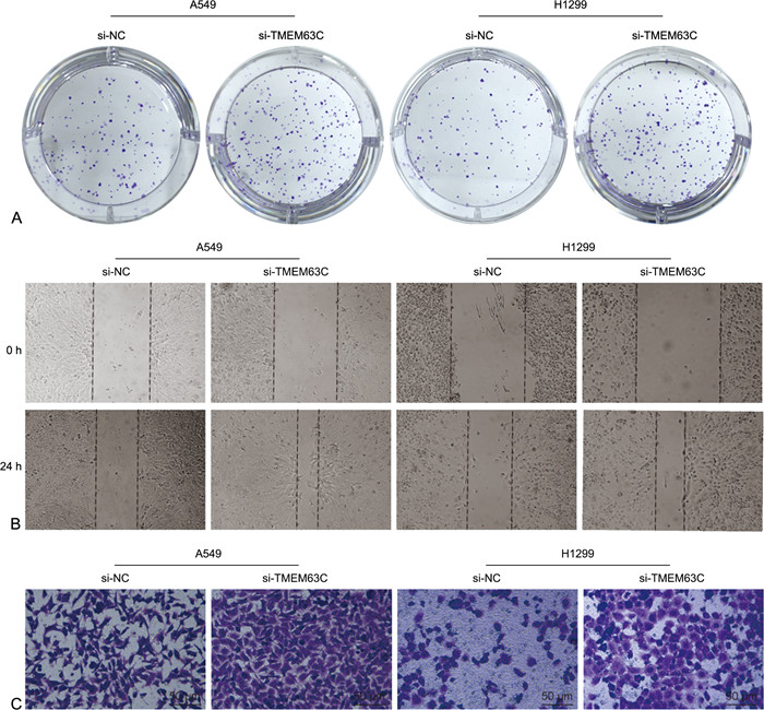
图 2 下调TMEM63C表达对LUAD细胞增殖、迁移与侵袭的影响
注:A为典型细胞克隆形成图片;B为典型细胞划痕与迁移图片;C为典型细胞Transwell侵袭图片。
Figure 2. Effects of TMEM63C knockdown on proliferation, migration, and invasion of LUAD cells

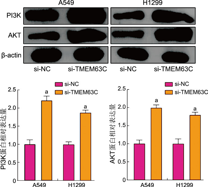
图 3 下调TMEM63C表达对LUAD细胞中PI3K、AKT蛋白表达的影响
注:与 si-NC 组比较,aP<0. 05。
Figure 3. Effects of TMEM63C knockdown on PI3K and AKT protein expression in LUAD cells
表 1 各组细胞中TMEM63C mRNA及蛋白相对表达量比较($\bar{x} \pm s$)
Table 1. Comparison of relative expression levels of TMEM63C mRNA and protein among cell groups ($\bar{x} \pm s$)
组别 n TMEM63C mRNA TMEM63C蛋白 BEAS-2B 3 1.00±0.03 1.00±0.13 A549 3 7.06±0.78a 2.12±0.16a H1299 3 10.96±0.43a 1.91±0.13a F值 288.400 53.840 P值 <0.001 <0.001 注:与BEAS-2B细胞系比较,aP<0.05。  下载: 导出CSV
表 2 基于TCGA-LUAD队列的单因素Cox回归分析
Table 2. Univariate Cox regression analysis based on the TCGA-LUAD cohort
变量 B SE Waldχ2 P值 HR(95% CI) TMEM63C mRNA -1.693 0.681 6.180 0.012 0.832(0.722~0.960) 年龄 -4.423 3.061 2.088 0.279 1.012(0.990~1.035) 性别(男性vs.女性) -2.323 1.796 1.673 0.648 1.103(0.724~1.679) TNM分期(Ⅱ期vs.Ⅰ期) 0.247 0.046 28.832 <0.001 3.597(2.130~6.078) TNM分期(Ⅲ期vs.Ⅰ期) 0.615 0.117 27.630 <0.001 6.350(3.669~10.986) TNM分期(Ⅳ期vs.Ⅰ期) 0.663 0.163 16.544 <0.001 6.976(3.150~15.442)
下载: 导出CSV
表 3 基于TCGA-LUAD队列的多因素Cox回归分析
Table 3. Multivariate Cox regression analysis based on the TCGA-LUAD cohort
变量 B SE Waldχ2 P值 HR(95% CI) TMEM63C mRNA -1.487 0.541 7.555 0.003 0.798(0.686~0.928) 年龄 4.135 2.621 2.489 0.155 1.016(0.994~1.039) 性别(男vs.女) -1.952 1.786 1.195 0.531 0.868(0.557~1.353) TNM分期(Ⅱ期vs.Ⅰ期) 0.182 0.046 15.654 <0.001 3.323(1.956~5.647) TNM分期(Ⅲ期vs.Ⅰ期) 0.582 0.117 24.744 <0.001 5.986(3.446~10.402) TNM分期(Ⅳ期vs.Ⅰ期) 0.525 0.163 10.374 <0.001 7.090(3.149~15.963) 注:各变量赋值方法如下,TMEM63C,低表达组=0, 高表达组=1;年龄<60岁=0, ≥60岁=1;女性=0, 男性=1;TNM分期,Ⅰ期=0, Ⅱ~Ⅳ期=1。
下载: 导出CSV
表 4 si-NC与si-TMEM63C组不同时间点A549细胞OD值比较($\bar{x} \pm s$)
Table 4. Comparison of OD values of A549 cells between si-NC and si-TMEM63C groups at different time points ($\bar{x} \pm s$)
组别 n 24 h 48 h 72 h 96 h si-NC 3 0.82±0.01 1.22±0.01a 2.30±0.11ab 3.08±0.06abc si-TMEM63C 3 0.81±0.02 1.80±0.08a 3.00±0.01ab 3.81±0.11abc t值 0.775 12.460 10.980 10.090 P值 0.482 <0.001 <0.001 <0.001 注:与24 h比较,aP<0.05;与48 h比较,bP<0.05;与72 h比较,cP<0.05。
下载: 导出CSV
表 5 si-NC与si-TMEM63C组不同时间点H1299细胞OD值比较($\bar{x} \pm s$)
Table 5. Comparison of OD values of H1299 cells between si-NC and si-TMEM63C groups at different time points ($\bar{x} \pm s$)
组别 n 24 h 48 h 72 h 96 h si-NC 3 0.70±0.01 1.00±0.01a 1.99±0.11ab 2.81±0.06abc si-TMEM63C 3 0.70±0.02 1.57±0.08a 1.57±0.08ab 3.55±0.11abc t值 0.001 12.250 5.348 10.230 P值 0.999 <0.001 0.006 <0.001 注:与24 h比较,aP<0.05;与48 h比较,bP<0.05;与72 h比较,cP<0.05。
下载: 导出CSV
表 6 si-NC与si-TMEM63C组A549细胞增殖、迁移与侵袭能力比较($\bar{x} \pm s$)
Table 6. Comparison of proliferation, migration, and invasion capacities of A549 cells between si-NC and si-TMEM63C groups ($\bar{x} \pm s$)
组别 n 克隆形成率(%) 细胞迁移率(%) 细胞侵袭数(个) si-NC 3 30.80±1.44 32.49±0.84 267.30±13.58 si-TMEM63C 3 49.33±3.63 59.72±2.03 373.00±25.87 t值 8.217 21.460 6.266 P值 0.001 <0.001 0.003
下载: 导出CSV
表 7 si-NC与si-TMEM63C组H1299细胞增殖、迁移与侵袭能力比较($\bar{x} \pm s$)
Table 7. Comparison of proliferation, migration, and invasion capacities of H1299 cells between si-NC and si-TMEM63C groups ($\bar{x} \pm s$)
组别 n 克隆形成率(%) 细胞迁移率(%) 细胞侵袭数(个) si-NC 3 24.73±2.55 35.55±1.15 80.33±1.16 si-TMEM63C 3 47.33±1.89 58.48±0.80 216.00±11.79 t值 12.340 28.370 19.840 P值 <0.001 <0.001 <0.001 注:与si-NC组比较,aP<0.05。
下载: 导出CSV
-
[1] BRAY F, LAVERSANNE M, SUNG H, et al. Global cancer statistics 2022: GLOBOCAN estimates of incidence and mortality worldwide for 36 cancers in 185 countries[J]. CA Cancer J Clin, 2024, 74(3): 229-263. [2] 黄庭, 贡会源, 唐震, 等. KLHL7在非小细胞肺癌中的表达及其临床意义[J]. 中华全科医学, 2023, 21(7): 1109-1112.HUANG T, GONG H Y, TANG Z, et al. Expression of KLHL7 in non-small cell lung cancer and its clinical significance[J]. Chinese Journal of General Practice, 2023, 21(7): 1109-1112. [3] SONG Y A, KELAVA L, KISS I. MiRNAs in lung adenocarcinoma: role, diagnosis, prognosis, and therapy[J]. Int J Mol Sci, 2023, 24(17): 13302. DOI: 10.3390/ijms241713302. [4] LIN H R, ZHANG X, FENG Y L, et al. Advancing lung adenocarcinoma prognosis and immunotherapy prediction with a multi-omics consensus machine learning approach[J]. J Cell Mol Med, 2024, 28(13): e18520. DOI: 10.1111/jcmm.18520. [5] KANG H, LEE C J. Transmembrane proteins with unknown function (TMEMs) as ion channels: electrophysiological properties, structure, and pathophysiological roles[J]. Exp Mol Med, 2024, 56(4): 850-860. doi: 10.1038/s12276-024-01206-1 [6] HERRERA-QUITERIO G A, ENCARNACIÓN-GUEVARA S. The transmembrane proteins (TMEM) and their role in cell proliferation, migration, invasion, and epithelial-mesenchymal transition in cancer[J]. Front Oncol, 2023, 13: 1244740. DOI: 10.3389/fonc.2023.1244740. [7] SCHMIT K, MICHIELS C. TMEM proteins in cancer: a review[J]. Front Pharmacol, 2018, 9: 1345. DOI: 10.3389/fphar.2018.01345. [8] 古联, 梁宝云, 周文君, 等. 跨膜蛋白63C基因遗传多态性与缺血性脑卒中气虚证显著关联[J]. 中国老年学杂志, 2023, 43(1): 15-20.GU L, LIANG B Y, ZHOU W J, et al. Transmembrane protein 63C gene polymorphism significantly associated with Qi deficiency syndrome of ischemic stroke[J]. Chinese Journal of Gerontology, 2023, 43(1): 15-20. [9] 朱明雪, 闫清, 王鹏, 等. EGFR突变对肺腺癌预后及治疗意义生物信息分析[J]. 青岛大学学报(医学版), 2023, 59(4): 522-526.ZHU M X, YAN Q, WANG P, et al. Significance of epidermal growth factor receptor mutation in the prognosis and treatment of lung adenocarcinoma: a bioinformatics analysis[J]. Journal of Qingdao University(Medical Sciences), 2023, 59(4): 522-526. [10] TÁBARA L C, AL-SALMI F, MAROOFIAN R, et al. TMEM63C mutations cause mitochondrial morphology defects and underlie hereditary spastic paraplegia[J]. Brain, 2022, 145(9): 3095-3107. doi: 10.1093/brain/awac123 [11] DE VISSER K E, JOYCE J A. The evolving tumor microenvironment: from cancer initiation to metastatic outgrowth[J]. Cancer Cell, 2023, 41(3): 374-403. [12] MYERS J A, MILLER J S. Exploring the NK cell platform for cancer immunotherapy[J]. Nat Rev Clin Oncol, 2021, 18(2): 85-100. doi: 10.1038/s41571-020-0426-7 [13] DEL PRETE A, SALVI V, SORIANI A, et al. Dendritic cell subsets in cancer immunity and tumor antigen sensing[J]. Cell Mol Immunol, 2023, 20(5): 432-447. [14] PLESCA I, MVLLER L, BÖTCHER J P, et al. Tumor-associated human dendritic cell subsets: phenotype, functional orientation, and clinical relevance[J]. Eur J Immunol, 2022, 52(11): 1750-1758. doi: 10.1002/eji.202149487 [15] XIONG S M, DONG L L, CHENG L. Neutrophils in cancer carcinogenesis and metastasis[J]. J Hematol Oncol, 2021, 14(1): 173. doi: 10.1186/s13045-021-01187-y [16] HE Y, SUN M M, ZHANG G G, et al. Targeting PI3K/Akt signal transduction for cancer therapy[J]. Signal Transduct Target Ther, 2021, 6(1): 425. -

 点击查看大图
计量
- 文章访问数: 12
- HTML全文浏览量: 8
- PDF下载量: 2
- 被引次数: 0
