



Diagnostic value of pelvic floor ultrasound and diastasis recti assessment in the evaluating pelvic floor function and postpartum stress urinary incontinence
-
摘要:
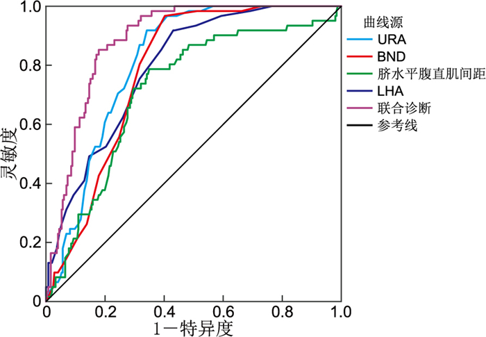
目的 探讨盆底超声参数及腹直肌分离程度与产后盆底功能障碍的关系,并分析其对压力性尿失禁(SUI)的诊断价值。 方法 回顾性分析2022年2月—2024年10月于安徽医科大学妇幼医学中心就诊的307例顺产产后女性,收集盆底超声最大Valsalva动作下的膀胱颈移动度(BND)、尿道旋转角(URA)、肛提肌裂孔面积(LHA)及膀胱后角(BA)、腹直肌间距(脐上3 cm、脐水平、脐下3 cm)。根据门诊病历查阅是否出现SUI相关症状,将其分为61例SUI组以及246例非SUI组,比较2组的差异,并通过logistic回归及ROC曲线评估指标的独立及联合诊断价值。 结果 SUI组BND、URA、LHA高于非SUI组,差异有统计学意义(P<0.001)。SUI组脐水平腹直肌间距高于非SUI组, 差异有统计学意义(P<0.001)。而2组的BA、脐上3 cm与脐下3 cm腹直肌间距差异无统计学意义(P>0.05)。经多因素logistic回归分析显示,BND、URA、LHA以及脐水平腹直肌间距均是产后发生SUI的影响因素(P<0.05)。ROC曲线结果显示,BND、URA、LHA以及脐水平腹直肌间距诊断产后SUI的曲线下面积分别为0.794、0.814、0.782、0.716(P<0.001),联合诊断曲线下面积可达0.886。 结论 产妇盆底超声指标、脐水平腹直肌间距对SUI具有一定的临床诊断价值。 Abstract:Objective To explore the relationship between pelvic floor ultrasound parameters and the degree of rectus abdominis diastasis in postpartum women, and to analyze their diagnostic value for stress urinary incontinence (SUI). Methods A retrospective analysis was performed on 307 postpartum women who had undergone vaginal delivery and attended the Maternal and Child Health Center, Anhui Medical University from February 2022 to October 2024. The bladder neck mobility (BND), urethral rotation angle (URA), latissimus dorsi muscle cleavage area (LHA), and bladder posterior angle (BA), as well as rectus abdominis muscle spacing measured during maximal Valsalva maneuver (at the umbilical level, 3 cm above the umbilicus, and 3 cm below the umbilicus), were collected. According to the outpatient medical records, participants were divided into 61 cases of SUI group and 246 cases of non-SUI group. Differences between the two groups were compared, and the independent and combined diagnostic values of the indexes were evaluated by multivariate logistic regression and ROC curve analyses. Results The BND, URA, and LHA were significantly higher in the SUI group than in the non-SUI group (P < 0.001). The rectus abdominis muscle spacing at the umbilical level was also higher in the SUI group than in the non-SUI group (P < 0.001). No statistically significant differences were observed in BA, 3 cm above the umbilicus, or 3 cm below the umbilicus (P> 0.05). After multifactorial logistic regression analysis identified BND, URA, LHA, and rectus abdominis muscle spacing at the umbilical level as independent influencing factors for postnatal SUI (P < 0.05). ROC curves showed that the areas under the curve for BND, URA, LHA, and rectus abdominis muscle spacing at the umbilical level were 0.794, 0.814, 0.782, and 0.716, respectively (P < 0.001), while the combined area under the curve could reached 0.886. Conclusion Maternal pelvic floor ultrasound indexes and abdominis muscle spacing at the umbilical level demonstrate clinical diagnostic value for the diagnosis of SUI. -

图 1 盆底超声指标、脐水平腹直肌间距诊断产后SUI的ROC曲线
Figure 1. ROC curve of pelvic floor ultrasound indices and umbilicus-level rectus abdominis diastasis for diagnosing postpartum stress urinary incontinence
表 1 2组产妇一般资料比较
Table 1. Comparison of general characteristics between two groups of postpartum women
项目 SUI组(n=61) 非SUI组(n=246) 统计量 P值 年龄(x±s,岁) 30.02±3.27 29.15±3.43 1.781a 0.076 孕次[M(P25, P75),次] 1(1, 2) 1(1, 2) -0.865b 0.387 产次[M(P25, P75),次] 1(1, 2) 1(1, 1) -1.051b 0.293 阴道手术助产[例(%)] 4(6.6) 9(3.7) 0.424c 0.515 身高[M(P25, P75),cm] 162.0(158.5, 167.0) 161.0(158.0, 165.0) -1.359b 0.174 孕前体重(x±s,kg) 56.91±7.68 55.17±7.74 1.576a 0.116 孕前BMI(x±s) 21.61±2.49 21.28±2.77 0.847a 0.398 孕周(x±s周) 39.63±0.90 39.64±1.01 0.091a 0.928 注: a为t值,b为Z值,c为χ2值。  下载: 导出CSV
表 2 2组产妇最大Valsalva动作下盆底超声相关指标比较(x±s)
Table 2. Comparison of pelvic floor ultrasound indicators during maximal Valsalva maneuver between two groups of postpartum women (x±s)
组别 例数 BND(mm) URA(°) BA(°) LHA(cm2) SUI组 61 30.54±5.73 68.26±10.20 115.43±18.95 26.61±2.82 非SUI组 246 23.80±6.23 53.26±14.45 110.91±21.20 22.33±4.85 t值 7.674 9.383 1.519 8.999 P值 <0.001 <0.001 0.130 <0.001
下载: 导出CSV
表 3 2组产妇静息状态下不同位置腹直肌间距比较(x±s)
Table 3. Comparison of inter-recti distance at different positions in the resting state between two groups of postpartum women (x±s)
组别 例数 腹直肌间距(cm) 脐上3 cm 脐水平 脐下3 cm SUI组 61 2.20±0.79 2.86±0.72 1.02±0.70 非SUI组 246 2.17±0.68 2.33±0.68 0.94±0.60 t值 0.310 5.461 0.850 P值 0.756 <0.001 0.422
下载: 导出CSV
表 4 影响产后发生SUI的多因素logistic回归分析
Table 4. Multivariate logistic regression analysis of factors associated with postpartum stress urinary incontinence
变量 B SE Waldχ2 P值 OR值 95% CI BND 0.133 0.033 16.013 <0.001 1.142 1.070~1.219 URA 0.042 0.014 8.671 0.003 1.043 1.014~1.073 LHA 0.153 0.050 9.404 0.002 1.165 1.057~1.285 脐水平腹直肌间距 0.751 0.245 9.408 0.002 2.119 1.311~3.425
下载: 导出CSV
表 5 盆底超声指标、脐水平腹直肌间距对产后SUI的诊断价值
Table 5. Diagnostic value of pelvic floor ultrasound indicators and umbilical-level inter-recti distance for postpartum stress urinary incontinence
项目 AUC SE 95% CI 灵敏度 特异度 约登指数 cut-off值 P值 BND(mm) 0.794 0.028 0.740~0.849 0.852 0.610 0.462 25.50 <0.001 URA(°) 0.814 0.024 0.767~0.861 0.918 0.659 0.577 55.50 <0.001 LHA(cm2) 0.782 0.026 0.731~0.833 0.967 0.598 0.565 23.50 <0.001 脐水平腹直肌间距(cm) 0.716 0.036 0.646~0.786 0.787 0.650 0.437 2.52 <0.001 联合 0.886 0.019 0.849~0.923 0.852 0.822 0.674 <0.001
下载: 导出CSV
-
[1] STROEDER R, RADOSA J, CLEMENS L, et al. Urogynecology in obstetrics: impact of pregnancy and delivery on pelvic floor disorders, a prospective longitudinal observational pilot study[J]. Arch Gynecol Obstet, 2021, 304(2): 401-408. doi: 10.1007/s00404-021-06022-w [2] CAVALLI M, AIOLFI A, BRUNI P G, et al. Prevalence and risk factors for diastasis recti abdominis: a review and proposal of a new anatomical variation[J]. Hernia, 2021, 25(4): 883-890. doi: 10.1007/s10029-021-02468-8 [3] YOU C, ZHAO Y J, ZHANG C, et al. Pelvic floor parameters predict postpartum stress urinary incontinence: a prospective MRI study[J]. Insights Imag, 2023, 14(1): 160. DOI: 10.1186/s13244-023-01488-5. [4] LIU X H, WANG Q, CHEN Y L, et al. Factors associated with stress urinary incontinence and diastasis of rectus abdominis in women at 6-8 weeks postpartum[J]. Urogynecology (Phila), 2023, 29(10): 844-850. doi: 10.1097/SPV.0000000000001353 [5] GONZÁLEZ-TIMONEDA A, VALLES-MURCIA N, MUÑOZ ESTEBAN P, et al. Prevalence and impact of pelvic floor dysfunctions on quality of life in women 5-10 years after their first vaginal or caesarian delivery[J]. Heliyon, 2025, 11(3): e42018. DOI: 10.1016/j.heliyon.2025.e42018. [6] 刘闪闪, 方悦, 周一波. 盆底超声评估女性混合性尿失禁疗效的临床研究[J]. 中华全科医学, 2025, 23(1): 107-110.LIU S S, FANG Y, ZHOU Y B. Study on the pelvic floor ultrasound for evaluating the therapeutic effect level of mixed urinary incontinence in women[J]. Chinese Journal of General Practice, 2025, 23(1): 107-110. [7] 李多, 鲁蓉. 盆底超声评估女性肛提肌形态及功能的研究进展[J]. 中南大学学报(医学版), 2023, 48(8): 1267-1273.LI D, LU R. Research progress in pelvic floor ultrasound for assessing the morphology and function of levator ani muscle in women[J]. Journal of Central South University (Medical Science), 2023, 48(8): 1267-1273. [8] 张小航, 张凤娟, 全丽娟, 等. 三维盆底超声参数与不同BMI阴道分娩产妇产后盆底功能的关系及对SUI的预测价值[J]. 中国妇幼保健, 2024, 39(20): 4103-4106.ZHANG X H, ZHANG F J, et al. NOT_FOUND[J]. Maternal and Child Health Care of China, 2024, 39(20): 4103-4106. [9] SKOURA A, BILLIS E, PAPANIKOLAOU D T, et al. Diastasis recti abdominis rehabilitation in the postpartum period: a scoping review of current clinical practice[J]. Int Urogynecol J, 2024, 35(3): 491-520. doi: 10.1007/s00192-024-05727-1 [10] THABET A A, ALSHEHRI M A. Efficacy of deep core stability exercise program in postpartum women with diastasis recti abdominis: a randomised controlled trial[J]. J Musculoskelet Neuronal Interact, 2019, 19(1): 62-68. [11] 中华医学会妇产科学分会妇科盆底学组. 女性压力性尿失禁诊断和治疗指南(2017)[J]. 中华妇产科杂志, 2017, 52(5): 289-293.Pelvic Floor Disease Group, Obstetrics and Gynecology Branch of Chinese Medical Association. NOT_FOUND[J]. Chinese Journal of Obstetrics and Gynecology, 2017, 52(5): 289-293. [12] 孙世甜, 殷伟红. 盆底超声诊断产后早期压力性尿失禁的研究进展[J]. 山东医药, 2022, 62(19): 109-112.SUN S T, YIN W H. Research progress of pelvic floor ultrasound in the diagnosis of early postpartum stress urinary incontinence[J]. Shandong Medical Journal, 2022, 62(19): 109-112. [13] DELANCEY J O L, MASTELING M, PIPITONE F, et al. Pelvic floor injury during vaginal birth is life-altering and preventable: what can we do about it?[J]. Am J Obstet Gynecol, 2024, 230(3): 279-294. e272. doi: 10.1016/j.ajog.2023.11.1253 [14] 吴凤姬, 陈梅. 经会阴三维超声测量肛提肌裂孔面积及形态诊断产后子宫脱垂的价值[J]. 现代医用影像学, 2024, 33(6): 1099-1102.WU F J, CHEN M. The Value of Measuring the Hiatus Area and Shape of Levator Anal Muscle by Three-Dimensional Perineal Ultrasonography in the Diagnosis of Postpartum Uterine Prolapse[J]. Modern Medical Imaging, 2024, 33(6): 1099-1102. [15] WANG X X, ZHANG L, LU Y. Advances in the molecular pathogenesis and cell therapy of stress urinary incontinence[J]. Front Cell Dev Biol, 2023, 11: 1090386. DOI: 10.3389/fcell.2023.1090386. [16] MOHR S, RAIO L, GOBRECHT-KELLER U, et al. Postpartum urinary retention: what are the sequelae? A long-term study and review of the literature[J]. Int Urogynecol J, 2022, 33(6): 1601-1608. doi: 10.1007/s00192-021-05074-5 [17] LAFRAMBOISE F C, SCHLAFF R A, BARUTH M. Postpartum exercise intervention targeting diastasis recti abdominis[J]. Int J Exerc Sci, 2021, 14(3): 400-409. [18] GLUPPE S, ENGH M E, BØ K. What is the evidence for abdominal and pelvic floor muscle training to treat diastasis recti abdominis postpartum? A systematic review with meta-analysis[J]. Braz J Phys Ther, 2021, 25(6): 664-675. doi: 10.1016/j.bjpt.2021.06.006 [19] 刘禹彤, 姜伊娜, 王春光, 等. 超声评估经阴道分娩产妇肛提肌裂孔面积与腹直肌距离及其相关性研究[J]. 医学影像学杂志, 2023, 33(12): 2257-2260.LIU Y T, JIANG Y N, et al. NOT_FOUND[J]. Journal of Medical Imaging, 2023, 33(12): 2257-2260. -

 点击查看大图
计量
- 文章访问数: 4
- HTML全文浏览量: 2
- PDF下载量: 1
- 被引次数: 0
