



An experience in the diagnosis and treatment of difficult-to-treat complicated urinary tract infection
-
摘要: 患者, 女性,29岁。临床以反复发热、腰痛、脓尿和肾区包块为主要表现,影像学提示右肾结构异常,呈“熊掌征”,存在鹿角形或长条形结石;肾血流功能显像提示右肾无功能。予充分抗感染治疗后患者仍间断排脓尿,伴炎症指标轻度增加。复习文献及请专科医师会诊,最终制定了以根治性右肾切除术为主的治疗方案。组织病理确诊为黄色肉芽肿性肾盂肾炎(xanthogranulomatous pyelonephritis,XGP),明确了疾病诊断。6个月后随访预后好。本文总结XGP的诊断要点,旨在提高全科医生对XGP的认识,使XGP患者尽早得到诊断、治疗和康复。
-
关键词:
- 黄色肉芽肿性肾盂肾炎 /
- 脓尿 /
- 熊掌肾
Abstract: A 29-year-old female presented with recurrent fever, lumbar pain, pyuria, and renal masses. Imaging revealed structural abnormalities of the right kidney, demonstrating the classical "Bear's Paw sign", with angular or linear calculi. Renal hemodynamic scintigraphy suggested that the right kidney was non-functional. Despite adequate anti-infective treatment, the patient continued to exhibit persistent pyuria with mildly increased inflammatory markers. Reviewing the literature and consulting multidisciplinary specialists, a therapeutic regimen centered on radical right nephrectomy was ultimately determined. Histopathology identified xanthogranulomatous pyelonephritis (XGP), establishing the final diagnosis. The prognosis of XGP was improved over a 6-month follow-up period. This study summarizes the key points of XGP diagnosis to improve the general physicians' awareness, facilitating earlier diagnosis, treatment, and rehabilitation.-
Key words:
- Xanthogranulomatous pyelonephritis /
- Pyuria /
- Bear's paw kidney
-

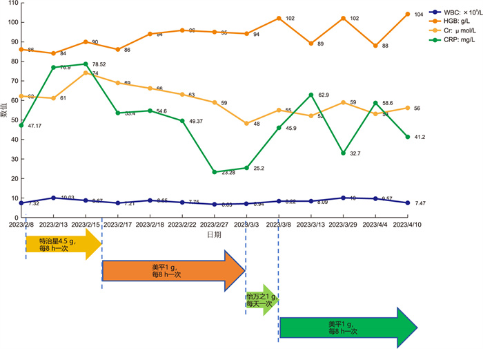
图 1 患者血WBC、Hb、Cr以及CRP变化
Figure 1. Changes in blood WBC, Hb, Cr, and CRP of the patient

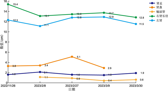
图 2 患者泌尿系结构参数变化
Figure 2. Changes in urinary system structural parameters of the patient
-
[1] HARRISON W, GOUGH P, RAVEENTHIRAN S, et al. Paediatric xanthogranulomatous pyelonephritis with reno-psoas fistula, psoas abscess and migration of renal calculi[J]. Urol Case Rep, 2023, 51: 102607. DOI: 10.1016/j.eucr.2023.102607. [2] TREEZA D J, DHUA A K, KANDASAMY D, et al. Xanthogranulomatous pyelonephritis in neonate: presentation, management and outcome[J]. BMJ Case Rep, 2023, 16(11): e255258. DOI: 10.1136/bcr-2023-255258. [3] XU Z, CAI T, ZHANG X, et al. Xanthogranulomatous pyelonephritis infected with the Providencia stuartii: a case report and literature review[J]. BMC Nephrol, 2021, 22(1): 356. DOI: 10.1186/s12882-021-02565-x. [4] 刘亮. 黄色肉芽肿性肾盂肾炎的CT影像诊断及鉴别分析[J]. 影像研究与医学应用, 2019, 3(17): 85-86.LIU L. CT imaging diagnosis and differential analysis of xanthogranulomatous pyelonephritis[J]. Imaging Research and Medical Applications, 2019, 3(17): 85-86. [5] ROMETTI M, PATEL D, BRYCZKOWSKI C. Xanthogranulomatous pyelonephritis: case report[J]. J Emerg Med, 2024, 66(4): e534-e537. doi: 10.1016/j.jemermed.2023.11.021 [6] SINGH M, ETHIRAJ D, INDIRAN V, et al. Xanthogranulomatous pyelonephritis with calculus migration into the psoas abscess: an unusual complication[J]. Autops Case Rep, 2020, 11: e2020200. DOI: 10.4322/acr.2020.200. [7] 陈柯行, 穆毅, 陈攀, 等. 黄色肉芽肿性肾盂肾炎1例并文献复习[J]. 疑难病杂志, 2021, 20(3): 289-290.CHEN K X, MU Y, CHEN P, et al. Xanthogranulomatous pyelonephritis: a case report and literature review[J]. Chinese Journal of Difficult and Complicated Cases, 2021, 20(3): 289-290. [8] 卓涛, 叶敏, 张金伟, 等. 黄色肉芽肿性肾盂肾炎的临床诊治分析(附41例报告)[J]. 中华泌尿外科杂志, 2019, 40(8): 578-582.ZHUO T, YE M, ZHANG J W, et al. Clinical diagnosis and treatment analysis of xanthogranulomatous pyelonephritis: a report of 41 cases[J]. Chinese Journal of Urology, 2019, 40(8): 578-582. [9] TONKAZ G, TONKAZ M M, ERKAL T D, et al. Xanthogranulomatous pyelonephritis: "Bear's paw sign"[J]. Med Clin (Barc), 2024, S0025-7753(24)00025-3. DOI: 10.1016/j.medcli.2023.11.044. [10] 郑思强, 刘琳, 范神栋, 等. 尿路感染中常见病原菌及其特殊类型对抗菌药物的耐药情况[J]. 中华全科医学, 2023, 21(11): 1852-1855. doi: 10.16766/j.cnki.issn.1674-4152.003241ZHENG S Q, LIU L, FAN S D, et al. Drug resistance of common pathogenic bacteria and their special types in urinary tract infections[J]. Chinese Journal of General Practice, 2023, 21(11): 1852-1855. doi: 10.16766/j.cnki.issn.1674-4152.003241 [11] 刘晓霞, 翟曜耀, 卢再鸣. 黄色肉芽肿性肾盂肾炎的CT影像诊断及病理分析[J]. 临床放射学杂志, 2021, 40(5): 941-944.LIU X X, ZHAI Y Y, LU Z M. CT imaging diagnosis and pathological analysis of xanthogranulomatous pyelonephritis[J]. Journal of Clinical Radiology, 2021, 40(5): 941-944. [12] ALENAZI K A. Atypical clinical presentation of xanthogranulomatous pyelonephritis in a female child: a case report[J]. Cureus, 2024, 16(2): e53666. DOI: 10.7759/cureus.53666. [13] FRIED A, TUERK C, SCHIMA W, et al. Xanthogranulomatous pyelonephritis with staghorn calculus, acute gangrenous appendicitis and enterocolitis: a multidisciplinary challenge of kidney-preserving conservative therapy[J]. Curr Urol, 2015, 8(3): 162-165. doi: 10.1159/000365709 [14] UPASANI A, BARNACLE A, ROEBUCK D, et al. Combination of surgical drainage and renal artery embolization: an alternative treatment for xanthogranulomatous pyelonephritis[J]. Cardiovase Intervent Radio, 2017, 40(3): 470-473. doi: 10.1007/s00270-016-1522-z -

 下载:
下载:
 点击查看大图
图(3)
计量
- 文章访问数: 3
- HTML全文浏览量: 2
- PDF下载量: 0
- 被引次数: 0
