



Establishment of a stable PD-L1-overexpressing oral squamous carcinoma MOC-1 cell line and verification of its in vivo function
-
摘要:
目的 构建程序性死亡蛋白1配体(PD-L1)稳定过表达的小鼠口腔鳞状细胞(MOC-1)稳转细胞模型,并探究其对口腔鳞状细胞癌(OSCC)体内生物学行为的影响。 方法 通过分子克隆技术构建pCDH-PD-L1过表达质粒,经慢病毒包装感染MOC-1细胞,嘌呤霉素筛选获得稳定过表达PD-L1的细胞株。采用RT-qPCR和Western blotting验证PD-L1的表达;采用完全随机设计,使用随机数表法将10只小鼠分为过PD-L1表达组和对照组,每组5只,进行皮下荷瘤实验评估体内成瘤能力;利用免疫组织化学、免疫荧光检测肿瘤组织中PD-L1、Ki67表达及CD8+T细胞浸润与杀伤功能。 结果 成功构建pCDH-PD-L1重组质粒;RT-qPCR及Western blotting证实PD-L1过表达MOC-1细胞系构建成功。动物荷瘤实验显示,PD-L1过表达组瘤体体积和重量均显著大于对照组[(361.11±54.05)mm3 vs. (94.94±23.15)mm3、(124.8±34.8)g vs. (14.0±4.9)g,P<0.001]。免疫检测结果表明,过表达组肿瘤组织中PD-L1阳性率[(60.5±4.2)% vs. (20.8±2.8)%, P<0.001]、Ki67阳性细胞比例[(61.2±7.1)% vs. (30.4±5.5)%, P<0.001]均显著升高,而CD8+T细胞浸润数量显著减少[(5 023±961)个/mm2 vs. (8 702±1 006)个/mm2, P<0.001)],且杀伤能力降低。 结论 本研究基于pCDH载体成功构建PD-L1过表达的体内外研究模型,PD-L1过表达可通过抑制CD8+T细胞浸润及杀伤能力,显著增强OSCC细胞的体内成瘤能力。 -
关键词:
- 口腔鳞状细胞癌 /
- 程序性死亡蛋白1配体 /
- 稳转细胞株 /
- 皮下荷瘤
Abstract:Objective To construct a stable MOC-1 cell line with constitutive overexpression of programmed death-ligand 1 (PD-L1) and to investigate its effects on the in vivo biological behaviors of oral squamous cell carcinoma (OSCC). Methods The pCDH-PD-L1 overexpression plasmid was constructed via molecular cloning techniques, and then packaged into lentiviruses to infect murine oral squamous cell carcinoma (MOC-1) cells. Stable transfectants were selected with puromycin. PD-L1 expression at both the mRNA and protein levels was validated by RT-qPCR and Western blot analysis. Using a completely randomized design, 10 mice were divided into a PD-L1 overexpression group and a control group (5 mice per group) using a random number table. Stable cells were then subcutaneously implanted into mice to evaluate in vivo tumorigenicity. Immunohistochemistry (IHC) and immunofluorescence (IF) staining were performed to assess the expression of PD-L1 and Ki67, as well as the infiltration and cytotoxic function of CD8+ T cells in tumor tissues. Results The pCDH-PD-L1 recombinant plasmid was successfully constructed. RT-qPCR and Western blot analyses confirmed the successful establishment of a stable PD-L1-overexpressing MOC-1 cell line. In vivo subcutaneous tumor experiments showed that PD-L1 overexpression significantly accelerated tumor growth, resulting in markedly larger tumor volume and weight compared with the control group [(361.11±54.05) mm3 vs. (94.94±23.15) mm3, (14.0±4.9) g vs. (124.8±34.8) g, P < 0.001]. Immunological analyses indicated that the positive rate of PD-L1 [(60.5±4.2) % vs. (20.8±2.8) %, P < 0.001] and the proportion of Ki67-positive cells [(61.2±7.1) % vs. (30.4±5.5) %, P < 0.001] in tumor tissues of the overexpression group were both significantly increased. In contrast, the infiltration number of CD8+ T cells was significantly reduced [(5 023±961) cells/mm2 vs. (8 702±1 006) cells/mm2, P < 0.001], accompanied by impaired cytotoxic capacity. Conclusion We successfully established in vitro and in vivo models of PD-L1 overexpression based on the pCDH vector. PD-L1 overexpression significantly enhances the in vivo tumorigenicity of OSCC cells by inhibiting the infiltration and cytotoxic capacity of CD8+ T cells. -

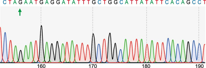
图 1 PD-L1过表达重组质粒测序结果
Figure 1. Sequencing results of PD-L1 overexpression recombinant plasmid

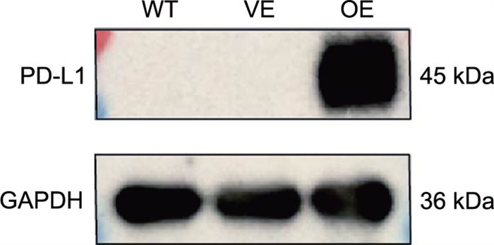
图 2 Western blotting验证稳转细胞株中PD-L1表达
Figure 2. Western blotting verified PD-L1 expression in stable cell lines

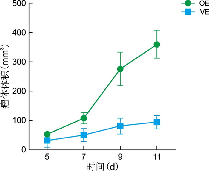
图 3 OE组和VE组小鼠皮下荷瘤瘤体体积比较
注:2组瘤体体积比较,aP<0.001。
Figure 3. Comparison of tumor volume of subcutaneous tumor-bearing in OE and VE groups

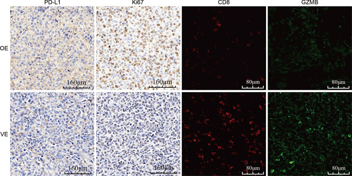
图 4 OE组和VE组小鼠免疫组化及免疫荧光结果(×40)
Figure 4. Results of immunohistochemistry and immunofluorescence in OE and VE groups (×40)
表 1 2组小鼠皮下荷瘤瘤体体积比较(x±s,mm3)
组别 只数 接种后第5天 接种后第7天 接种后第9天 接种后第11天 OE组 5 53.10±5.10 108.10±14.35 276.60±51.60 361.11±54.05 VE组 5 32.05±21.07 51.21±20.59 81.20±20.45 94. 94±23.15 F值 0.797 2.153 7.397 10.080 P值 0.991 0.425 <0.001 <0.001 注:F组间=39.830,P<0.001;F时间=128.500,P<0.001;F交互=17.950,P<0.001。  下载: 导出CSV
-
[1] ALQUTUB S, ALQUTUB A, BAKHSHWIN A, et al. Histopathological predictors of lymph node metastasis in oral cavity squamous cell carcinoma: a systematic review and meta-analysis[J]. Front Oncol, 2024, 14: 1401211. DOI: 10.3389/fonc.2024.1401211. [2] XUE N N, WANG Y, WANG Z Y, et al. Trends in immunotherapy for oral squamous cell carcinoma[J]. Cell Oncol(Dordr), 2025, 48(5): 1159-1179. doi: 10.1007/s13402-025-01068-3 [3] CHENG Y X, SONG Z Z, CHEN J, et al. Molecular basis, potential biomarkers, and future prospects of OSCC and PD-1/PD-L1 related immunotherapy methods[J]. Heliyon, 2024, 10(4): e25895. DOI: 10.1016/j.heliyon.2024.e25895. [4] KUJAN O, AGAG M, SMAGA M, et al. PD-1/PD-L1, Treg-related proteins, and tumour-infiltrating lymphocytes are associated with the development of oral squamous cell carcinoma[J]. Pathology, 2022, 54(4): 409-416. doi: 10.1016/j.pathol.2021.09.013 [5] XU S B, WANG M Y, SHI X Z, et al. Influence of PD-1/PD-L1 on immune microenvironment in oral leukoplakia and oral squamous cell carcinoma[J]. Oral Dis, 2023, 29(8): 3268-3277. doi: 10.1111/odi.14332 [6] TANG Q, CHEN Y, LI X J, et al. The role of PD-1/PD-L1 and application of immune-checkpoint inhibitors in human cancers[J]. Front Immunol, 2022, 13: 964442. DOI: 10.3389/fimmu.2022.964442. [7] KUJAN O, VAN SCHAIJIK B, FARAH C S. Immune checkpoint inhibitors in oral cavity squamous cell carcinoma and oral potentially malignant disorders: a systematic review[J]. Cancers, 2020, 12(7): 1937. DOI: 10.3390/cancers12071937. [8] MU H R, ZHANG Q, ZUO D Q, et al. Methionine intervention induces PD-L1 expression to enhance the immune checkpoint therapy response in MTAP-deleted osteosarcoma[J]. Cell Rep Med, 2025, 6(3): 101977. DOI: 10.1016/j.xcrm.2025.101977. [9] GAO Y, FENG Y F, LIU S C, et al. Immune-independent acquired resistance to PD-L1 antibody initiated by PD-L1 upregulation via PI3K/AKT signaling can be reversed by anlotinib[J]. Cancer Med, 2023, 12(14): 15337-15349. doi: 10.1002/cam4.6195 [10] 陈志红, 吴亚东. 程序性死亡受体-1及其配体在口腔鳞状细胞癌中的研究进展[J]. 华西口腔医学杂志, 2020, 38(4): 449-453.Chen Z H, Wu D Y. Development of programmed death receptor-1 and programmed death receptor-1 ligand in oral squamous cell carcinoma[J]. West China Journal of Stomatology, 2020, 38(4): 449-453. [11] ZHOU Y J, LI G L, WANG J Y, et al. PD-L1: expression regulation[J]. Blood Sci, 2023, 5(2): 77-91. doi: 10.1097/BS9.0000000000000149 [12] YANG Z, LIU X P, ZHU J, et al. Inhibiting intracellular CD28 in cancer cells enhances antitumor immunity and overcomes anti-PD-1 resistance via targeting PD-L1[J]. Cancer Cell, 2025, 43(1): 86-102. e10. doi: 10.1016/j.ccell.2024.11.008 [13] 房恩岳, 张丽萍, 马雪征, 等. 慢病毒载体系统及其安全性研究进展[J]. 病毒学报, 2023, 39(4): 1181-1192.Fang E Y, Zhang L P, Ma X Z, et al. Research progress on lentiviral vector systems and their safety issue[J]. Chinese Journal of Virology, 2023, 39(4): 1181-1192. [14] GARCIA-DIAZ A, SHIN D S, MORENO B H, et al. Interferon receptor signaling pathways regulating PD-L1 and PD-L2 expression[J]. Cell Rep, 2017, 19(6): 1189-1201. doi: 10.1016/j.celrep.2017.04.031 [15] JUNEJA V R, MCGUIRE K A, MANGUSO R T, et al. PD-L1 on tumor cells is sufficient for immune evasion in immunogenic tumors and inhibits CD8 T cell cytotoxicity[J]. J Exp Med, 2017, 214(4): 895-904. doi: 10.1084/jem.20160801 [16] 孙奥龙, 张丽柯, 徐全晓, 等. 非小细胞肺癌患者中GZMB和PD-L1的表达及两者与预后的关系[J]. 中华全科医学, 2024, 22(11): 1863-1867. doi: 10.16766/j.cnki.issn.1674-4152.003751Sun A L, Zhang L K, Xu Q X, et al. The expression of GZMB and PD-L1 and their relationship with prognosis in non-small cell lung patients[J]. Chinese Journal of General Practice, 2024, 22(11): 1863-1867. doi: 10.16766/j.cnki.issn.1674-4152.003751
 点击查看大图
计量
- 文章访问数: 5
- HTML全文浏览量: 3
- PDF下载量: 0
- 被引次数: 0
