



Application of shear wave elastography in perineal assessment during vaginal delivery
-
摘要:
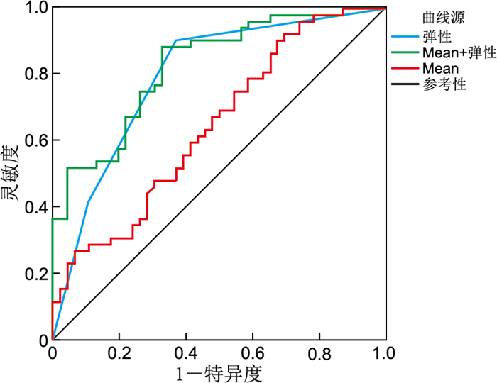
目的 应用剪切波弹性成像技术(SWE)对晚孕女性会阴体组织进行定量测量,分析其评估会阴体弹性的效能, 探讨其在阴道分娩临床会阴评估中的应用价值。 方法 采用前瞻性研究设计,便利抽样选取2022年3月—2023年11月在安徽医科大学第一附属医院进行产前检查符合纳入标准的197例晚孕女性,采用剪切波弹性成像技术测量其会阴体弹性。随诊其分娩结局并收集阴道分娩产妇临床会阴评估指标数据,剔除产钳助产、第二产程延长和急性胎儿窘迫病例后,以阴道分娩时是否行会阴切开将观察对象分为会阴切开组(53例)和非会阴切开组(50例),比较2组的会阴评估指标。以会阴切开为因变量,进行logistic回归分析,构建联合预测模型,绘制ROC曲线,分析SWE测量数据对会阴切开的预测价值。 结果 会阴切开组与非会阴切开组临床会阴评估指标中会阴体弹性差异有统计学意义(P < 0.05)。2组SWE测量结果中Max、Min和Mean值比较差异均有统计学意义(P < 0.05)。采用Mean值、弹性及两者联合3种方法评估会阴是否需要切开的AUC(95% CI)分别为0.647(0.538~0.756)、0.796(0.705~0.887)、0.829(0.749~0.908),灵敏度分别为0.904、0.904、0.885,特异度分别为0.674、0.630、0.674,约登指数分别为0.230、0.534、0.559,Mean联合弹性的预测效能优于单独预测。 结论 临床会阴体弹性评估联合SWE测量可提供更加准确的评估效能,显示出良好的应用前景。 Abstract:Objective To employ shear wave elastography (SWE) technology to quantitatively measure the perineal body tissue in late-pregnant women, analyze its efficacy in assessing perineal body elasticity, and explore its application value in the clinical perineal assessment for vaginal delivery. Methods A prospective study design was employed. Convenience sampling was employed to select 197 late-pregnant women who met the inclusion criteria and underwent prenatal examinations at the First Affiliated Hospital of Anhui Medical University from March 2022 to November 2023. SWE was used to measure perineal body elasticity. Their delivery outcomes were followed up, and clinical perineal assessment indicators were collected for those who underwent vaginal delivery. After excluding cases involving forceps-assisted delivery, prolonged second stage of labor, and acute fetal distress, participants were categorized into an episiotomy group (53 cases) and a non-episiotomy group (50 cases) based on whether an episiotomy was performed during vaginal delivery. The perineal assessment indicators were compared between the two groups. Using episiotomy as the dependent variable, logistic regression was performed, a combined prediction model was constructed, and a receiver operating characteristic (ROC) curve was plotted to analyze the predictive value of SWE measurements for episiotomy. Results Among the clinical perineal assessment parameters, perineal body elasticity showed a statistically significant difference (P < 0.05) between the episiotomy and non-episiotomy groups. There were statistically significant differences (P < 0.05) in the Max, Min, and Mean values of SWE measurements between the two groups. The areas under the ROC curve (AUC, 95% CI) for predicting the necessity of episiotomy using SWE Mean value, clinical elasticity assessment, and their combination were 0.647 (0.538-0.756), 0.796 (0.705-0.887), and 0.829 (0.749-0.908), respectively. The corresponding sensitivities were 0.904, 0.904, and 0.885; specificities were 0.674, 0.630, and 0.674; and Youden indices were 0.230, 0.534, and 0.559. The predictive efficacy of the Mean value combined with clinical elasticity was superior to that of either clinical elasticity or the Mean value alone. Conclusion Clinical perineal body elasticity assessment in combination with SWE measurement offers enhanced assessment accuracy and shows favorable application prospects. -
Key words:
- Shear wave elastography /
- Perineal body /
- Vaginal delivery /
- Perineal assessment
-

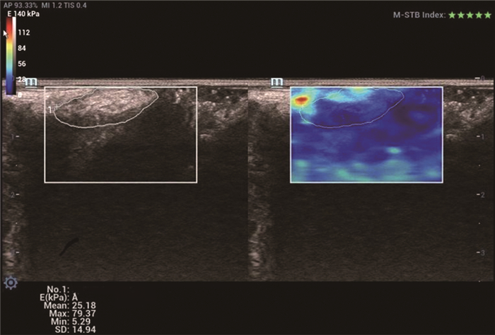
图 1 妊娠≥36周孕妇会阴体剪切波弹性成像
Figure 1. Shear wave elastography of the perineal body in pregnant women at ≥36 weeks of gestation

图 2 Mean、弹性、Mean+弹性评估会阴体侧切的ROC曲线
Figure 2. ROC Curves for Mean, Elasticity, and Mean + Elasticity in assessing the need for lateral episiotomy
表 1 会阴切开组与非会阴切开组产妇的一般资料比较
Table 1. Comparison of general data between parturients with and without episiotomy
组别 例数 年龄(x±s,岁) 产前BMI (x±s) 孕前BMI [M(P25, P75)] 分娩孕天[M(P25, P75),d] 新生儿体质量(x±s,g) 非会阴切开组 50 29.14±3.78 27.24±2.91 20.85(19.27, 23.40) 282.00(277.75, 287.00) 3 412.20±393.93 会阴切开组 53 28.60±3.32 27.33±3.72 21.30(19.45, 23.55) 283.00(277.50, 286.00) 3 352.64±227.57 统计量 0.763a 0.131a -0.178b -0.076b 0.921a P值 0.447 0.896 0.859 0.939 0.359 注:a为t值,b为Z值。  下载: 导出CSV
表 2 会阴切开组与非会阴切开组产妇的临床会阴评估指标和SWE参数比较
Table 2. Comparison of clinical perineal assessment indicators and swe parameters between parturients with and without episiotomy
项目 非会阴切开组(n=50) 会阴切开组(n=53) 统计量 P值 会阴体弹性(例) -5.365a <0.001 差 5 22 一般 12 25 好 29 5 会阴体长度(例) 1.050b 0.305 <3 cm 9 14 3~7 cm 41 39 会阴体水肿(例) 4.204b 0.122 无 46 41 轻微 1 9 有 3 3 Max[M(P25, P75),kPa] 74.01(64.25, 85.18) 85.70(71.25, 93.61) -2.531a 0.011 Min[M(P25, P75),kPa] 8.60(6.75, 11.82) 10.75(8.20, 18.33) -2.597a 0.009 Mean(x±s,kPa) 30.62±7.59 34.87±6.64 3.014c 0.003 SD(x±s,kPa) 12.11±2.78 13.36±3.19 2.118c 0.037 注:a为Z值,b为χ2值,c为t值。会阴体弹性非会阴切开组缺失4例,会阴切开组缺失1例。
下载: 导出CSV
表 3 产妇会阴侧切影响因素的logistic回归分析
Table 3. Logistic regression analysis of influencing factors of lateral episiotomy in parturients
变量 B SE Waldχ2 P值 OR值 95% CI Mean 0.083 0.037 5.130 0.024 1.087 1.011~1.167 会阴体弹性 1.696 0.374 20.567 <0.001 5.453 2.620~11.349
下载: 导出CSV
表 4 3种会阴评估指标对会阴切开的评估效能比较
Table 4. Comparison of the efficacy of three perineal assessment indicators for episiotomy
项目 AUC(95% CI) 截断值(kPa) 灵敏度 特异度 约登指数 Mean 0.647(0.538~0.756) 26.065 0.904 0.674 0.230 弹性 0.796(0.705~0.887) 0.904 0.630 0.534 Mean+弹性 0.829(0.749~0.908) 0.885 0.674 0.559
下载: 导出CSV
-
[1] RADNER G, JENNEWEIN L, BRÜGGMANN D, et al. The impact of selective episiotomy on maternal short-term morbidity: a retrospective study[J]. J Obstet Gynaecol, 2024, 44(1): 2369664. DOI: 10.1080/01443615.2024.2369664. [2] 张旱梅, 周利华, 马红, 等. 会阴切开的发生率及影响因素分析[J]. 中华全科医学, 2022, 20(9): 1517-1520, 1572. doi: 10.16766/j.cnki.issn.1674-4152.002638Zhang H M, Zhou L H, Ma H, et al. Incidence and influencing factors of perineotomy[J]. Chinese General Practice Journal, 2022, 20(9): 1517-1520, 1572. doi: 10.16766/j.cnki.issn.1674-4152.002638 [3] HU T T, ZHAO L H, ZHAO X L, et al. Accurate prediction of mediolateral episiotomy risk during labor: development and verification of an artificial intelligence model[J]. BMC Pregnancy Childbirth, 2025, 25(1): 370. DOI: 10.1186/s12884-025-07441-2. [4] 徐佳丹, 周双双. 会阴评估量表在产妇分娩中会阴切开的应用效果分析[J]. 浙江创伤外科, 2024, 29(6): 1056-1058.Xu J D, Zhou S S. Analysis of the application effect of perineal assessment scale in episiotomy during childbirth[J]. Zhejiang Journal of Traumatic Surgery, 2024, 29(6): 1056-1058. [5] 朱弘, 阮玲欢. 会阴切开评估表在限制会阴切开中的应用[J]. 中国临床护理, 2023, 15(11): 685-688.Zhu H, Ruan L H. Application of perineotomy evaluation table in limiting perineotomy[J]. Chinese Journal of Clinical Nursing, 2023, 15(11): 685-688. [6] CHEN L Y, LOW L K, DELANCEY J O, et al. In vivo estimation of perineal body properties using ultrasound quasistatic elastography in nulliparous women[J]. J Biomech, 2015, 48(9): 1575-1579. doi: 10.1016/j.jbiomech.2015.02.056 [7] GACHON B, FRITEL X, PIERRE F, et al. In vivo assessment of the elastic properties of women's pelvic floor during pregnancy using shear wave elastography: design and protocol of the ELASTOPELV study[J]. BMC Musculoskelet disord, 2020, 21(1): 305. DOI: 10.1186/s12891-020-03333-y. [8] 黎婷, 陆永萍, 杨寒凝, 等. 实时剪切波超声弹性成像技术评估健康女性肛门括约肌复合体的初步研究[J]. 中国临床医学影像杂志, 2024, 35(7): 484-489.Li T, Lu Y P, Yang H N, et al. A preliminary study of real-time shear wave ultrasonic elastography to evaluate the anal sphincter complex in healthy women[J]. Chinese Journal of Clinical Medical Imaging, 2024, 35(7): 484-489. [9] 周敏知. 女性会阴体形态学及其对盆底支持功能的超声影像学研究[D]. 上海: 上海交通大学, 2020.Zhou M Z. Ultrasonographic study on female perineal body morphology and its role in pelvic floor support function[D]. Shanghai: Shanghai Jiao Tong University, 2022. [10] 李彦, 付丽曼, 李娜, 等. 经会阴超声实时剪切波弹性成像预测早产风险的价值[J]. 中国医学影像学杂志, 2022, 30(12): 1267-1271.Li Y, Fu L M, Li N, et al. Application of real-time shear wave elastography by using transperineal ultrasound for predicting preterm labor in pregnant women[J]. Chinese Journal of Medical Imaging, 2022, 30(12): 1267-1271. [11] 朱雯, 马桂凤, 林秀梅, 等. SWE联合灰阶超声评估不同分度子宫脱垂初产妇会阴体变化[J]. 医学影像学杂志, 2021, 31(2): 302-305.Zhu W, Ma G F, Lin X M, et al. SWE combined with gray-scale ultrasound to evaluate perineal changes in primiparas with different degrees of uterine prolapse[J]. Journal of Medical Imaging, 2021, 31(2): 302-305. [12] 董丽丽, 王冰霜, 李晓静, 等. 超声剪切波弹性成像联合盆底超声检查对阴道分娩产妇盆底功能障碍的诊断效能分析[J]. 中国医刊, 2023, 58(10): 1130-1134.Dong L L, Wang B S, Li X J, et al. Diagnostic efficacy of shear wave elastography combined with pelvic floor ultrasound in pelvic floor dysfunction in women after vaginal delivery[J]. Chinese Journal of Medicine, 2023, 58(10): 1130-1134. [13] 李刚, 徐华勤, 薛超, 等. 经会阴盆底超声联合剪切波弹性成像对阴道分娩产妇盆腔器官脱垂的诊断价值[J]. 临床超声医学杂志, 2023, 25(9): 713-717.Li G, Xu H Q, Xue C, et al. Value of transperineal pelvic floor ultrasound combined with shear wave elastography in the diagnosis of pelvic organ prolapse in puerpera with vaginal delivery[J]. Journal of Clinical Ultrasound in Medicine, 2023, 25(9): 713-717. [14] ABE-TAKAHASHI Y, KITTA T, OUCHI M, et al. Morphological examination of pelvic floor muscles in a rat model of vaginal delivery[J]. BMC Pregnancy Childbirth, 2024, 24(1): 95. DOI: 10.1186/s12884-024-06278-5. [15] DURAN P, BOSCOLO SESILLO F, COOK M, et al. Proregenerative extracellular matrix hydrogel mitigates pathological alterations of pelvic skeletal muscles after birth injury[J]. Sci Transl Med, 2023, 15(707): eabj3138. DOI: 10.1126/scitranslmed.abj3138. [16] LI X M, ZHANG L M, LI Y, et al. Assessment of perineal body properties in women with stress urinary incontinence using Transperineal shear wave elastography[J]. Sci Rep, 2024, 14(1): 21647. DOI: 10.1038/s41598-024-72429-5.
 点击查看大图
计量
- 文章访问数: 3
- HTML全文浏览量: 2
- PDF下载量: 0
- 被引次数: 0
