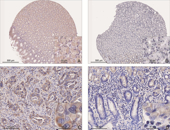
Expression and clinical significance of ALDH8A1 in gastric cancer tissues
-
摘要:
目的 探究重组表达醛脱氢酶8A1(ALDH8A1)在胃癌组织中的表达情况及与临床病理参数的关系,并分析其对患者远期预后的影响。 方法 纳入2012年3月—2018年4月在蚌埠医科大学第一附属医院接受根治术治疗的109例胃癌患者,采用免疫组化法分析ALDH8A1在胃癌及癌旁组织的表达情况,并运用统计学方法分析其与临床病理特征的关系;采用Kaplan-Meier曲线分析ALDH8A1的表达对胃癌患者术后5年生存率的影响。 结果 ALDH8A1在胃癌组织中的表达明显高于癌旁组织(P<0.001)。CEA≥5 μg/L、CA19-9≥37 kU/L、癌细胞类型为腺癌、T3~4期及N2~3期患者ALDH8A1表达水平更高(P<0.05)。Kaplan-Meier曲线分析及多因素分析显示ALDH8A1高表达(HR=2.674,95% CI:1.293~5.533,P=0.008)可能会增加胃癌患者术后的死亡率。ROC曲线表明以2.555为截点值,ALDH8A1对胃癌患者预后具有较高的预判价值(灵敏度为68.97%,特异度为86.27%)。GO和KEGG富集分析显示ALDH8A1与细胞减数分裂、DNA结合和细胞周期等信号通路有关。 结论 ALDH8A1在胃癌组织中高表达并促进胃癌的恶性进展,与预后不良相关。 -
关键词:
- 胃癌 /
- 重组表达醛脱氢酶8A1 /
- 临床病理特征 /
- 预后
Abstract:Objective To investigate the expression of aldehyde dehydrogenase 8 family member A1 (ALDH8A1) in gastric cancer tissues and its relationship with clinicopathological parameters, and to analyze its effect on the long-term prognosis of patients. Methods A total of 109 patients with gastric cancer who underwent radical resection in the First Affiliated Hospital of Bengbu Medical University from March 2012 to April 2018 were enrolled. Immunohistochemistry was used to analyze the expression of ALDH8A1 in gastric cancer tissues and adjacent tissues, and statistical methods were used to analyze its relationship with clinicopathological features. Kaplan-Meier Plotter curve was used to analyze the effect of ALDH8A1 expression on the 5-year survival rate of patients with gastric cancer after surgery. Results The expression of ALDH8A1 in gastric cancer tissues was significantly higher than that in adjacent tissues (P < 0.001). The high expression rate of ALDH8A1 was higher in patients with CEA≥5 μg/L, CA19-9≥37 kU/L, adenocarcinoma, T3-4 stage and N2-3 stage (P < 0.05). Kaplan-Meier Plotter curve analysis and multivariate analysis showed that high ALDH8A1 expression (HR=2.674, 95% CI: 1.293-5.533, P=0.008) may increase the mortality of patients with gastric cancer after surgery. ROC curve showed that with 2.555 as the cut-off value, ALDH8A1 had a high predictive value for the prognosis of gastric cancer patients (sensitivity was 68.97%, specificity was 86.27%). GO and KEGG enrichment analysis showed that ALDH8A1 was related to cell meiosis, DNA binding and cell cycle signaling pathways. Conclusion ALDH8A1 is highly expressed in gastric cancer tissues and promotes the malignant progression of gastric cancer and is associated with poor prognosis. -
表 1 胃癌患者ALDH8A1表达水平与临床病理参数的关系[例(%)]
Table 1. Relationship between ALDH8A1 expression level and clinicopathological parameters in gastric cancer patients[cases(%)]
项目 例数 ALDH8A1 χ2值 P值 低表达(n=55) 高表达(n=54) 性别 0.074 0.785 男性 78 40(51.28) 38(48.72) 女性 31 15(48.39) 16(51.61) 年龄(岁) 0.860 0.354 <60 39 22(56.41) 17(43.59) ≥60 70 33(47.14) 37(52.86) CEA(μg/L) 11.246 0.001 <5 54 36(66.67) 18(33.33) ≥5 55 19(34.55) 36(65.45) CA19-9(kU/L) 22.581 < 0.001 <37 47 36(76.60) 11(23.40) ≥37 62 19(30.65) 43(69.35) 肿瘤直径(cm) 1.614 0.204 <5 47 27(57.45) 20(42.55) ≥5 62 28(45.16) 34(54.84) 组织学分型 4.975 0.026 腺癌 61 25(40.98) 36(59.02) 其他 48 30(62.50) 18(37.50) T分期 11.245 0.001 1~2期 58 38(65.52) 20(34.48) 3~4期 51 17(33.33) 34(66.67) N分期 6.694 0.010 0~1期 54 34(62.96) 20(37.04) 2~3期 55 21(38.18) 34(61.82) 表 2 胃癌患者术后5年生存率影响因素的单因素及多因素分析
Table 2. Univariate and multivariate analysis of factors influencing the 5-year postoperative survival rate in gastric cancer patients
变量 单因素分析 多因素分析 log-rank χ2 P值 B SE Waldχ2 P值 HR值 95% CI 性别 1.406 0.236 年龄 0.615 0.433 ALDH8A1表达 35.350 < 0.001 0.984 0.371 7.034 0.008 2.674 1.293~5.533 CEA 25.434 < 0.001 0.878 0.339 6.727 0.009 2.406 1.239~4.672 CA19-9 31.379 < 0.001 1.314 0.405 10.527 0.001 3.721 1.683~8.231 肿瘤直径 0.928 0.335 组织学分型 2.271 0.132 T分期 22.032 < 0.001 0.693 0.319 4.723 0.030 2.000 1.070~3.737 N分期 17.594 < 0.001 0.941 0.336 7.849 0.005 2.562 1.327~4.946 -
[1] SUNG H, FERLAY J, SIEGEL R L, et al. Global cancer statistics 2020: GLOBOCAN estimates of incidence and mortality worldwide for 36 cancers in 185 countries[J]. CA Cancer J Clin, 2021, 71(3): 209-249. doi: 10.3322/caac.21660 [2] AJANI J A, D' AMICO T A, BENTREM D J, et al. Gastric cancer, version 2.2022, NCCN clinical practice guidelines in oncology[J]. J Natl Compr Canc Netw, 2022, 20(2): 167-192. doi: 10.6004/jnccn.2022.0008 [3] 曹芝郡, 姜永珊, 李吉彦, 等. 胃癌前病变细胞模型的建立及其在中医药研究中的应用[J]. 中国中医基础医学杂志, 2023, 29(7): 1208-1212. https://www.cnki.com.cn/Article/CJFDTOTAL-ZYJC202307034.htmCAO Z J, JIANG Y S, LI J Y, et al. Establishment of cell model for gastric precancerous lesions and its application in traditional chinese medicine research[J]. Journal Of Basic Chinese Medicine, 2023, 29(7): 1208-1212. https://www.cnki.com.cn/Article/CJFDTOTAL-ZYJC202307034.htm [4] 黄亮, 李昌伟, 余壮明. 胃癌患者癌组织EphA5表达与临床病理特征的关系[J]. 中华实用诊断与治疗杂志, 2023, 37(8): 781-786. https://www.cnki.com.cn/Article/CJFDTOTAL-HNZD202308006.htmHUANG L, LI C W, YU Z M. Correlations of EphA5 expression in gastric cancer tissues with clinicopathological features[J]. Journal of Chinese Practical Diagnosis and Therapy, 2023, 37(8): 781-786. https://www.cnki.com.cn/Article/CJFDTOTAL-HNZD202308006.htm [5] 韩亮, 张德龙, 桑卫东, 等. FBLN1 mRNA在胃腺癌组织中的表达及其临床意义[J]. 北京医学, 2020, 42(8): 720-723, 727. https://www.cnki.com.cn/Article/CJFDTOTAL-BJYX202008006.htmHAN L, ZHANG D L, SANG W D, et al. Expression of FBLN1 mRNA in stomach adenocarcinoma tissues and its clinical significance[J]. Beijing Medical Journal, 2020, 42(8): 720-723, 727. https://www.cnki.com.cn/Article/CJFDTOTAL-BJYX202008006.htm [6] 何旭旭, 赵萌, 杨一群, 等. UGT8在胃癌组织中的表达及对胃癌细胞转移的调控作用[J]. 中华全科医学, 2023, 21(4): 544-548. doi: 10.16766/j.cnki.issn.1674-4152.002927HE X X, ZHAO M, YANG Y Q, et al. UGT8 expression in gastric cancer tissues and its regulatory effect on gastric cancer metastasis[J]. Chinese Journal of General Practice, 2023, 21(4): 544-548. doi: 10.16766/j.cnki.issn.1674-4152.002927 [7] 牛云霞, 蒙锦莹, 韩林育. 胃癌组织中MARCKS、HIF-1α的表达水平与临床病理特征的关系[J]. 海南医学, 2023, 34(22): 3200-3204. doi: 10.3969/j.issn.1003-6350.2023.22.002NIU Y X, MENG J Y, HAN L Y. Relationship between expression levels of MARCKS and HIF-1αin gastric cancer tissues and clinicopathological characteristics[J]. Hainan Medical Journal, 2023, 34(22): 3200-3204. doi: 10.3969/j.issn.1003-6350.2023.22.002 [8] 邓小婧, 辜正策, 李璐池, 等. 胃癌组织中lncRNA CHRF、miR-489的表达变化及意义[J]. 山东医药, 2023, 63(35): 51-54. doi: 10.3969/j.issn.1002-266X.2023.35.011DENG X J, GU Z C, LI L C, et al. Expression changes and significance of lncRNA CHRF and miR-489 in gastric cancer tissues[J]. Shandong Medical Journal, 2023, 63(35): 51-54. doi: 10.3969/j.issn.1002-266X.2023.35.011 [9] 黄莎莎, 鞠瑛, 段文冰, 等. C反应蛋白与白蛋白比值在早期胃癌辅助诊断中的价值[J]. 中国医药, 2022, 17(9): 1366-1370. https://www.cnki.com.cn/Article/CJFDTOTAL-ZGYG202209020.htmHUANG S S, JU Y, DUAN W B, et al. The value of C-reactive protein to albumin ratio in the auxiliary diagnosis of early gastric cancer[J]. China Medicine, 2022, 17(9): 1366-1370. https://www.cnki.com.cn/Article/CJFDTOTAL-ZGYG202209020.htm [10] 梁思远, 韦传毅, 龙子雯, 等. 术前外周血血小板/淋巴细胞比值相关列线图模型预测胃癌患者术后无病生存期: 一项双中心回顾性研究[J]. 中国临床医学, 2023, 30(3): 391-396. https://www.cnki.com.cn/Article/CJFDTOTAL-LCYX202303001.htmLIANG S Y, WEI C Y, LONG Z W, et al. Value of a preoperative peripheral platelet-to-lymphocyte ratio ralated nomogram model predicting postoperative disease-free survival in patients with gastric cancer: a two-center retrospective study[J]. Chinese Journal of Clinical Medicine, 2023, 30(3): 391-396. https://www.cnki.com.cn/Article/CJFDTOTAL-LCYX202303001.htm [11] 徐娇, 邹永红, 王敏. 术前血清ALT/AST值对早期胃癌患者术后复发的预测价值[J]. 医学临床研究, 2023, 40(3): 356-358, 362. doi: 10.3969/j.issn.1671-7171.2023.03.010XU J, ZOU Y H, WANG M. Predictive value of preoperative serum ALT/AST ratio for postoperative recurrence of early gastric cance[J]. Journal of Clinical Research, 2023, 40(3): 356-358, 362. doi: 10.3969/j.issn.1671-7171.2023.03.010 [12] ZHANG T, WANG B F, SU F, et al. TCF7L2 promotes anoikis resistance and metastasis of gastric cancer by transcriptionally activating PLAUR[J]. Int J Biol Sci, 2022, 18(11): 4560-4577. doi: 10.7150/ijbs.69933 [13] SHI M S, GU Y, JIN K F, et al. CD47 expression in gastric cancer clinical correlates and association with macrophage infiltration[J]. Cancer Immunol Immunother, 2021, 70(7): 1831-1840. doi: 10.1007/s00262-020-02806-2 [14] ZHOU X, YANG J W, LU Y Q, et al. Relationships of tumor differentiation and immune infiltration in gastric cancers revealed by single-cell RNA-seq analyses[J]. Cell Mol Life Sci, 2023, 80(2): 57. DOI: 10.1007/s00018-023-04702-1. [15] XIA J, LI S Q, LIU S L, et al. Aldehyde dehydrogenase in solid tumors and other diseases: potential biomarkers and therapeutic targets[J]. MedComm (2020), 2023, 4(1): e195. DOI: 10.1002/mco2.195. [16] HE J F, ZHAO H C, DENG D F, et al. Screening of significant biomarkers related with prognosis of liver cancer by lncRNA-associated ceRNAs analysis[J]. J Cell Physiol, 2020, 235(3): 2464-2477. doi: 10.1002/jcp.29151 [17] THRIFT A P, EL-SERAG H B. Burden of gastric cancer[J]. Clin Gastroenterol Hepatol, 2020, 18(3): 534-542. doi: 10.1016/j.cgh.2019.07.045 [18] KIM J, BAE D H, KIM J H, et al. HOXC10 overexpression promotes cell proliferation and migration in gastric cancer[J]. Oncol Rep, 2019, 42(1): 202-212. [19] JINAWATH N, SHIAO M S, CHANPANITKITCHOTE P, et al. Enhancement of migration and invasion of gastric cancer cells by IQGAP3 [J]. Biomolecules, 2020, 10(8): 1194. DOI: 10.3390/biom10081194. [20] TIAN S, PENG P L, LI J, et al. SERPINH1 regulates EMT and gastric cancer metastasis via the Wnt/β-catenin signaling pathway[J]. Aging (Albany NY), 2020, 12(4): 3574-3593. [21] 王金乐, 朱星蓉, 夏爱丹, 等. 京尼平苷调控Wnt/β-cantenin信号通路抑制胃癌转移的机制研究[J]. 浙江临床医学, 2022, 24(6): 810-813. https://www.cnki.com.cn/Article/CJFDTOTAL-LICX202206005.htmWANG J L, ZHU X R, XIA A D, et al. The mechanism research of Geniposide regulating Wnt/β-cantenin signaling pathway to inhibit gastric cancer metastasis[J]. Zhejiang Clinical Medical Journal, 2022, 24(6): 810-813. https://www.cnki.com.cn/Article/CJFDTOTAL-LICX202206005.htm [22] WANG X B, ZHAO Y J, LUO J, et al. MicroRNA hsa-miR-1301-3p regulates human ADH6, ALDH5A1 and ALDH8A1 in the ethanol-acetaldehyde-acetate metabolic pathway[J]. Mol Pharmacol, 2020, 98(2): 120-129. doi: 10.1124/mol.120.119693 -


 下载:
下载:


