Value of combined exploration of carotid plaque by multiple ultrasound techniques to predict recurrence of ischemic stroke
-
摘要:
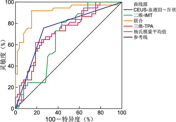
目的 探讨二维和三维超声、超声造影(CEUS)和剪切波弹性成像(SWE)联合探查颈动脉斑块并分析其预测缺血性脑卒中(IS)复发的价值。 方法 筛选新疆医科大学第二附属医院2021年10月—2023年1月收治的首发IS合并颈动脉粥样硬化斑块患者95例,根据是否复发分为复发组和未复发组。采用logistic回归分析研究影响IS患者复发的危险因素。采用ROC曲线分析多项超声技术联合预测IS复发的价值。 结果 失访5例,有37例患者复发,复发率为41.11%,复发组高血压占比高于未复发组,药物依从性好占比则低于未复发组(P<0.05)。复发组CEUS-血流Ⅲ~Ⅳ级占比及二维-动脉内中膜厚度(IMT)、三维-颈动脉斑块总面积(TPA)值高于未复发组,杨氏模量平均值则低于未复发组(P<0.05)。多因素logistic回归分析显示,CEUS-血流Ⅲ~Ⅳ级、二维-IMT高和三维-TPA大及杨氏模量平均值低是影响IS患者复发的危险因素,药物依从性好则是其保护因素(P<0.05)。ROC曲线分析显示,二维-IMT、三维-TPA、杨氏模量平均值及CEUS-血流Ⅲ~Ⅳ级单项预测IS患者复发的AUC分别为0.674、0.717、0.729、0.746,联合预测AUC为0.908,高于单项检测(P<0.05)。 结论 二维、三维及SWE、CEUS超声技术通过评估颈动脉斑块不稳定性,在预测IS复发方面具有一定的临床价值,四者联合价值更高。 Abstract:Objective To investigate the value of combined 2D and 3D ultrasound, contrast-enhanced ultrasonography (CEUS) and shear-wave elastography (SWE) for the detection of carotid plaques, and to analyze their value in predicting recurrence of ischaemic stroke (IS). Methods A total of 95 patients with first-ever IS and carotid atherosclerotic plaque from October, 2021 to January, 2023 in the Second Affiliated Hospital of Xinjiang Medical University were selected, and divided into a recurrence group and a non-recurrence group according to whether they had recurred or not. Risk factors for relapse in patients with IS were analyzed using logistic regression. Receiver operating characteristic (ROC) curves were used to analyze the value of different ultrasound techniques in predicting the recurrence of IS. Results Five cases were lost to follow-up. The recurrence rate was 41.11%, and there were no deaths after treatment. The proportion of hypertension in the recurrent group was higher than that in the non-recurrent group, and the proportion of good medication compliance was lower than that in the non-recurrent group (P < 0.05). The ratio of CEUS blood flow grade Ⅲ to Ⅳ, 2-D intima-media thickness (IMT) and 3-D-TPA in the recurrent group were higher than those in the non-recurrent, while the mean Young's modulus was lower than that in the non-recurrent group (P < 0.05). Multivariate logistic regression analysis showed that CEUS blood flow level Ⅲ to Ⅳ, high 2-IMT, large 3-3D total carotid plaque area (TPA) and low mean Young's modulus were risk factors for recurrence in IS patients, and good medication compliance was a protective factor (P < 0.05). ROC curve analysis showed that the AUC of 2-D IMT, 3-D TPA, mean Young's modulus and CEUS blood flow Ⅲ to Ⅳ in predicting recurrence of in IS patients were 0.674, 0.717, 0.729 and 0.746, respectively. The combined predictive AUC of 0.908 was higher than that of single detection (P < 0.05). Conclusion 2D, 3D and SWE and CEUS ultrasound techniques have some clinical value in predicting the recurrence of IS by evaluating carotid plaque instability, and the combined value of the four is higher. -
表 1 复发组与未复发组首发IS合并颈动脉粥样硬化斑块患者一般资料比较
Table 1. Comparison of general data between relapsed and non-relapsed groups in patients with first IS complicated with carotid atherosclerotic plaque
项目 复发组(n=37) 未复发组(n=53) 统计量 P值 性别(男/女,例) 20/17 23/30 0.992a 0.319 年龄(x±s,岁) 62.34±5.60 63.15±5.39 0.690b 0.492 BMI(x±s) 25.10±2.19 24.95±2.08 0.329b 0.743 吸烟史[例(%)] 8(21.62) 10(18.87) 0.103a 0.748 饮酒史[例(%)] 9(24.32) 11(20.75) 0.161a 0.689 合并症[例(%)] 糖尿病 9(24.32) 9(16.98) 0.734a 0.391 高血压 17(45.95) 10(18.87) 7.608a 0.006 高脂血症 12(32.43) 14(26.42) 0.384a 0.535 斑块侧别[例(%)] 1.537a 0.464 左侧 20(54.05) 25(47.17) 右侧 15(40.54) 21(39.62) 双侧 2(5.41) 7(13.21) 冠心病史[例(%)] 7(18.92) 6(11.32) 1.018a 0.313 脑卒中家族史[例(%)] 5(13.51) 5(9.43) 0.367a 0.545 药物依从性[例(%)] 12.274a <0.001 好 12(32.43) 37(69.81) 差 25(67.57) 16(30.19) NIHSS评分(x±s,分) 7.68±2.14 8.02±1.62 0.858b 0.393 注:a为χ2值,b为t值。 表 2 复发组与未复发组首发IS合并颈动脉粥样硬化斑块患者影像学参数比较
Table 2. Comparison of imaging parameters between recurrent and non-recurrent groups in patients with first IS and carotid atherosclerotic plaque
组别 例数 二维-IMT (x±s, mm) 三维-TPA (x±s, cm2) SWE(x±s, kPa) CEUS-血流分级[例(%)] 杨氏模量平均值 硬度分布标准差 Ⅰ~Ⅱ级 Ⅲ~Ⅳ级 复发组 37 1.67±0.39 22.06±3.20 42.33±9.12 21.92±5.45 9(24.32) 28(75.68) 未复发组 53 0.92±0.28 15.34±2.61 50.28±10.17 23.70±5.58 39(73.58) 14(26.42) 统计量 10.626a 10.945a 3.804a 1.503a 21.244b P值 <0.001 <0.001 <0.001 0.136 <0.001 注:a为t值,b为χ2值。 表 3 IS患者复发的多因素logistic回归分析
Table 3. Multivariate logistic regression analysis of relapse in patients with IS
变量 B SE Waldχ2 P值 OR值 95% CI 药物依从性好 -0.526 0.409 1.654 0.004 0.591 0.371~0.859 CEUS-血流Ⅲ~Ⅳ级 0.508 0.303 2.811 <0.001 1.662 1.336~2.874 二维-IMT 0.447 0.285 2.460 <0.001 1.564 1.148~2.652 三维-TPA 0.545 0.372 2.146 <0.001 1.725 1.309~3.001 杨氏模量平均值 0.472 0.298 2.509 <0.001 1.603 1.256~2.715 表 4 超声二维、三维及SWE、CEUS对IS患者复发的预测价值
Table 4. Predictive value of 2D, 3D ultrasound and SWE and CEUS for recurrence in patients with IS
指标 最佳截断值 灵敏度(%) 特异度(%) 约登指数 AUC 95% CI 二维-IMT 1.28 mm 81.08 56.60 0.379 0.674 0.567~0.769 三维-TPA 19.35 cm2 64.86 73.58 0.385 0.717 0.613~0.807 杨氏模量平均值 39.33 kPa 62.16 81.13 0.433 0.729 0.626~0.818 CEUS-血流Ⅲ~Ⅳ级 75.68 73.58 0.493 0.746 0.644~0.832 联合 91.89 84.91 0.768 0.908 0.828~0.959 -
[1] 《中国脑卒中防治报告》编写组. 《中国脑卒中防治报告2020》概要[J]. 中国脑血管病杂志, 2022, 19(2): 136-144. https://www.cnki.com.cn/Article/CJFDTOTAL-NXGB202311009.htmReport on stroke prevention and treatment in China Writing Group. Brief report on stroke prevention and treatment in China, 2020[J]. Chinese Journal of Cerebrovascular Diseases, 2019, 19(2): 136-144. https://www.cnki.com.cn/Article/CJFDTOTAL-NXGB202311009.htm [2] 中国心血管健康与疾病报告编写组. 中国心血管健康与疾病报告2022概要[J]. 中国循环杂志, 2023, 38(6): 583-612. https://www.cnki.com.cn/Article/CJFDTOTAL-ZGXH202407010.htmThe Writing Committee of the Report on Cardiovascular Health and Diseases in China. Report on cardiovascular health and diseases in China 2022: an updated summary[J]. Chinese Circulation Journal, 2023, 38(6): 583-612. https://www.cnki.com.cn/Article/CJFDTOTAL-ZGXH202407010.htm [3] 汤爱洁, 牛淑珍, 刘怡凡, 等. 基于倾向性评分匹配法评估急性缺血性脑卒中神经功能改善的影响因素分析[J]. 中华全科医学, 2022, 20(2): 186-189. doi: 10.16766/j.cnki.issn.1674-4152.002308TANG A J, NIU S Z, LIU Y F, et al. Analysis of the influencing factors of neurological improvement in acute ischemic stroke on the basis of propensity score matching[J]. Chinese Journal of General Practice, 2002, 20(2): 186-189. doi: 10.16766/j.cnki.issn.1674-4152.002308 [4] ZHOU F, HUA Y, JI X, et al. A systemic review into carotid plaque features as predictors of restenosis after carotid endarterectomy[J]. J Vasc Surg, 2021, 73(6): 2179-2188. [5] KASHIWAZAKI D, YAMAMOTO S, AKIOKA N, et al. Dilated microvessel with endothelial cell proliferation involves intraplaque hemorrhage in unstable carotid plaque[J]. Acta Neurochir (Wien), 2021, 163(6): 1777-1785. [6] ZHANG Y, CAO J, ZHOU J, et al. Plaque elasticity and intraplaque neovascularisation on carotid artery ultrasound: a comparative histological study[J]. Eur J Vasc Endovasc Surg, 2021, 62(3): 358-366. [7] ZAMANI M, SKAGEN K, SCOTT H, et al. Advanced ultrasound methods in assessment of carotid plaque instability: a prospective multimodal study[J]. BMC Neurol, 2020, 20(1): 39. [8] 中华医学会神经病学分会, 中华医学会神经病学分会脑血管病学组. 中国急性缺血性脑卒中诊治指南2018[J]. 中华神经科杂志, 2018, 51(9): 666-682. https://www.cnki.com.cn/Article/CJFDTOTAL-XDJB201911024.htmChinese Society of Neurology; Chinese Stroke Society. Chinese guidelines for diagnosis and treatment of acute ischemic stroke 2018[J]. Chinese Journal of Neurology, 2018, 51(9): 666-682. https://www.cnki.com.cn/Article/CJFDTOTAL-XDJB201911024.htm [9] 黄雅萍, 侯放, 文晟, 等. 超声造影诊断颈动脉不稳定斑块破裂风险的效能[J]. 中国超声医学杂志, 2023, 39(1): 5-8. https://www.cnki.com.cn/Article/CJFDTOTAL-ZGCY202301002.htmHUANG Y P, HOU F, WEN S, et al. Efficacy of contrast-enhanced ultrasound in diagnosing the risk of carotid artery unstable plaque rupture[J]. Chinese Journal of Ultrasound Medicine, 2019, 39(1): 5-8. https://www.cnki.com.cn/Article/CJFDTOTAL-ZGCY202301002.htm [10] CHALOS V, VAN DER ENDE N A M, LINGSMA H F, et al. National institutes of health stroke scale: an alternative primary outcome measure for trials of acute treatment for ischemic stroke[J]. Stroke, 2020, 51(1): 282-290. [11] SCHINKEL A F L, BOSCH J G, STAUB D, et al. Contrast-Enhanced ultrasound to assess carotid intraplaque neovascularizatione[J]. Ultrasound Med Biol, 2020, 46(3): 466-478. [12] $\overline{\mathrm{S}}$KOLOUDÍK D, KE$\overline{\mathrm{S}}$NEROVÁ P, VOMÁČKA J, et al. Shear-Wave elastography enables identification of unstable carotid plaque[J]. Ultrasound Med Biol, 2021, 47(7): 1704-1710. https://www.cnki.com.cn/Article/CJFDTOTAL-TJIY202404007.htm [13] KABŁAK-ZIEMBICKA A, PRZEWŁOCKI T. Clinical significance of carotid intima-media complex and carotid plaque assessment by ultrasound for the prediction of adverse cardiovascular events in primary and secondary care patients[J]. J Clin Med, 2021, 10(20): 4628. DOI: 10.3390/jcm10204628. [14] YOON H J, KIM K H, PARK H, et al. Carotid plaque rather than intima-media thickness as a predictor of recurrent vascular events in patients with acute ischemic stroke[J]. Cardiovasc Ultrasound, 2017, 15(1): 19. DOI: 10.1186/s12947-017-0110-y. [15] HENSLEY B, HUANG C, CRUZ MARTINEZ C V, et al. Ultrasound measurement of carotid intima-media thickness and plaques in predicting coronary artery disease[J]. Ultrasound Med Biol, 2020, 46(7): 1608-1613. [16] 张莉, 李安洋, 郭磊, 等. 颈动脉斑块彩色多普勒超声联合头颈部CTA诊断缺血性脑卒中患者颈动脉狭窄临床价值[J]. 医学影像学杂志, 2023, 33(8): 1477-1480. https://www.cnki.com.cn/Article/CJFDTOTAL-XYXZ202308036.htmZHANG L, LI A Y, GUO L, et al. Clinical value of color Doppler ultrasound combined with head and neck CTA in diagnosing carotid artery stenosis in patients with ischemic stroke[J]. Journal of Medical Imaging, 2023, 33(8): 1477-1480. https://www.cnki.com.cn/Article/CJFDTOTAL-XYXZ202308036.htm [17] ZHANG S, JIANG S, WANG C, et al. Comparison of ultrasonic shear wave elastography, AngioPLUS planewave ultrasensitive imaging, and optimized high-resolution magnetic resonance imaging in evaluating carotid plaque stability[J]. Peer J, 2023, 11: e16150. DOI: 10.7717/peerj.16150. [18] GOUDOT G, SITRUK J, JIMENEZ A, et al. Carotid plaque vulnerability assessed by combined shear wave elastography and ultrafast doppler compared to histology[J]. Transl Stroke Res, 2022, 13(1): 100-111. [19] 宫文亮, 周建, 朱道伟. 超声造影联合剪切波弹性成像评估乳腺癌病灶可切除性的研究[J]. 影像科学与光化学, 2022, 40(6): 1375-1380. https://www.cnki.com.cn/Article/CJFDTOTAL-GKGH202206011.htmGONG W L, ZHOU J, ZHU D W. Evaluation of resectability of breast cancer lesions by contrast-enhanced ultrasound combined with shear wave elastography[J]. Imaging Science and Photochemistry, 2022, 40(6): 1375-1380. https://www.cnki.com.cn/Article/CJFDTOTAL-GKGH202206011.htm [20] JOO S P, LEE S W, CHO Y H, et al. Vasa vasorum densities in human carotid atherosclerosis is associated with plaque development and vulnerability[J]. J Korean Neurosurg Soc, 2020, 63(2): 178-187. [21] CAMPS-RENOM P, PRATS-SÁNCHEZ L, CASONI F, et al. Plaque neovascularization detected with contrast-enhanced ultrasound predicts ischaemic stroke recurrence in patients with carotid atherosclerosis[J]. Eur J Neurol, 2020, 27(5): 809-816. [22] 刘燕霞, 郭林佳, 王俐元, 等. 北京市缺血性脑卒中复发高危患者健康信念及二级预防现状调查[J]. 中国社会医学杂志, 2022, 39(6): 671-675. https://www.cnki.com.cn/Article/CJFDTOTAL-GWSY202206015.htmLIU Y X, GUO L J, WANG L Y, et al. Investigation on health belief and secondary prevention status of patients at high risk of recurrent ischemic stroke in Beijing[J]. Chinese Journal of Social Medicine, 2022, 39(6): 671-675. https://www.cnki.com.cn/Article/CJFDTOTAL-GWSY202206015.htm -


 下载:
下载:


