

Evaluation effect and clinical application value of CT angiography on plaque status and luminal stenosis in patients with coronary heart disease
-
摘要:
目的 分析CT血管成像(CTA)对冠心病患者斑块状态、管腔狭窄的评估效果及临床应用价值,以期为临床寻找适宜的无创检查方法提供参考。 方法 回顾性分析郑州市第七人民医院2019年10月—2022年10月收治的166例冠心病患者的临床资料,以冠状动脉造影(CAG)为“金标准”,计算CTA与CAG检查结果的一致性。基于CTA比较不同狭窄程度患者的冠脉节段斑块类型,分析CTA冠脉斑块特征及狭窄程度对主要心脏不良事件(MACE)发生风险的影响。 结果 CAG共检出冠脉狭窄病变172处,CTA检出灵敏度、特异度及符合率分别为96.51%(166/172)、71.54%(352/492)、78.01%(518/664),与CAG结果的一致性良好(Kappa=0.818,P<0.001)。经CTA检查共检出89处冠脉斑块,轻度狭窄冠脉节段的非钙化(35.71%,10/28)、混合斑块(32.14%, 9/28)占比分别较中/重度狭窄[57.37%(35/61)、39.34%(24/61)]冠脉节段低,轻度狭窄冠脉节段钙化斑块(32.14%, 9/28)占比较中/重度狭窄(3.27%, 2/61)冠脉节段高,差异均有统计学意义(P<0.05)。非钙化斑块体积、斑块长度及管腔直径狭窄率是MACE发生的独立影响因素(P<0.001)。 结论 CTA可较为准确地评估冠心病患者斑块状态、管腔狭窄情况。 Abstract:Objective To evaluate the evaluation effect and clinical application value of CT angiography (CTA) on plaque status and lumen stenosis in patients with coronary heart disease, in order to provide reference for clinical search for appropriate non-invasive examination methods. Methods The clinical data of 166 patients with coronary heart disease admitted to the Seventh People' s Hospital of Zhengzhou from October 2019 to October 2022 were retrospectively analyzed. Coronary angiography (CAG) was used as the "gold standard" to calculate the consistency of CTA and CAG results. The types of coronary segmental plaque in patients with different degrees of stenosis were compared based on CTA, and the effects of coronary plaque characteristics and stenosis degree on the risk of major adverse cardiac events (MACE) were analyzed. Results A total of 172 coronary stenosis lesions were detected by CAG, and the sensitivity, specificity and coincidence rate of CTA were 96.51% (166/172), 71.54% (352/492) and 78.01% (518/664), respectively, which were in good agreement with the results of CAG (Kappa=0.818, P < 0.001). A total of 89 coronary plaques were detected by CTA. The proportions of non-calcified plaques (35.71%, 10/28) and mixed plaques (32.14%, 9/28) in the mild stenosis segments were lower than those in the moderate/severe stenosis segments [57.37%(35/61), 39.34%(24/61)]. The proportion of calcified plaques in mild stenosis segments (32.14%, 9/28) was higher than that in moderate/severe stenosis segments (3.27%, 2/61), and the difference was statistically significant (P < 0.05). Non-calcified plaque volume, plaque length and lumen diameter stenosis rate were independent risk factors for MACE (P < 0.001). Conclusion CTA can accurately evaluate the plaque status and lumen stenosis in patients with coronary heart disease. -
Key words:
- Coronary heart disease /
- Plaque status /
- CT angiography /
- Luminal stenosis
-

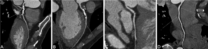
图 1 冠心病CTA图像
注:A为前降支钙化及非钙化斑块并管腔轻-中度狭窄,约50%;B为中段部分管腔走行于心肌内,回旋支钙化及非钙化斑块并管腔轻度狭窄,约25%;C为右冠状动脉钙化及非钙化斑块并管腔轻度狭窄,约30%;D为后降支非钙化斑块并管腔轻度狭窄,约30%,对角支、钝缘支、圆锥支、锐缘支管腔充盈良好,未见明显异常。
Figure 1. CTA image of coronary heart disease
表 1 CTA与CAG对冠脉狭窄病变的检出情况
Table 1. The detection of coronary artery stenosis lesions by CTA and CAG
CTA CAG 合计 病变血管节段 正常血管节段 病变血管节段 166 40 206 正常血管节段 6 352 358 合计 172 492 664  下载: 导出CSV
表 2 CTA与CAG诊断冠脉管腔狭窄程度的结果比较[例(%)]
Table 2. Comparison of the results of CTA and CAG in the diagnosis of coronary vessel lumen stenosis[cases(%)]
检查方法 例数 轻度狭窄 中度狭窄 重度狭窄 CAG 172 75(43.60) 69(40.12) 28(16.28) CTA 206 99(48.06) 63(30.58) 44(21.36) 注:2组冠脉管腔狭窄程度比较,Z=-1.825, P=0.068。
下载: 导出CSV
表 3 CTA检出钙化斑块在不同管腔狭窄中的分布情况[例(%)]
Table 3. Distribution of calcified plaque in different lumen stenoses detected by CTA[cases(%)]
管腔狭窄程度 例数 非钙化斑块 混合斑块 钙化斑块 轻度狭窄 28 10(35.71) 9(32.14) 9(32.14) 中/重度狭窄 61 35(57.37) 24(39.34) 2(3.27) 注:2组钙化斑块分布情况比较,χ2=14.986,P<0.001。
下载: 导出CSV
表 4 有、无MACE者CTA管腔狭窄及冠脉斑块特征比较(x±s)
Table 4. Comparison of CTA lumen stenosis and coronary plaque characteristics between patients with and without MACE(x±s)
组别 例数 非钙化斑块体积
(mm3)钙化斑块体积
(mm3)斑块负荷
(%)混合斑块体积
(mm3)管腔直径狭窄率
(%)MACE组 29 164.65±25.14 8.76±1.11 0.72±0.12 128.56±9.45 67.56±5.56 无MACE组 137 106.85±19.54 9.03±1.16 0.59±0.06 126.55±8.31 56.33±3.56 t值 13.724 1.147 5.685 1.155 10.434 P值 <0.001 0.253 <0.001 0.250 <0.001
下载: 导出CSV
表 5 变量赋值情况
Table 5. Variable assignment
变量 赋值方法 非钙化斑块体积 ≤136 mm3=0,>136 mm3=1 斑块负荷 ≤0.67%=0,>0.67%=1 管腔直径狭窄率 ≤60.5%=0,>60.5%=1
下载: 导出CSV
表 6 CTA管腔狭窄及冠脉斑块特征影响MACE发生的多因素分析
Table 6. Multivariate analysis of lumen stenosis and coronary plaque characteristics on CTA influencing the occurrence of MACE
变量 B SE Waldχ2 P值 OR(95% CI) 非钙化斑块体积 0.414 0.152 9.124 <0.001 1.513(1.123~2.038) 斑块负荷 0.577 0.214 11.574 <0.001 1.781(1.171~2.709) 管腔直径狭窄率 0.401 0.187 8.524 <0.001 1.493(1.035~2.154)
下载: 导出CSV
-
[1] 杨玲, 杜雪平. 社区规范管理的稳定性冠心病患者生活质量研究[J]. 中华全科医学, 2023, 21(11): 1898-1902. doi: 10.16766/j.cnki.issn.1674-4152.003253YANG L, DU X P. Research on the quality of life in patients with stable coronary artery disease under standardized management in community[J]. Chinese Journal of General Practice, 2023, 21(11): 1898-1902. doi: 10.16766/j.cnki.issn.1674-4152.003253 [2] RAJA J, SEITA M P, YEDLAPATI N, et al. Can computed fractional flow reserve coronary CT angiography (FFRCT) offer an accurate noninvasive comparison to invasive coronary angiography (ICA)?[J]. Curr Probl Cardiol, 2020, 46(3): 100642. DOI: 10.1016/j.cpcardiol.2020.100642. [3] 丁华永. CTA在冠心病冠脉狭窄程度及斑块状态评估中的应用[J]. 中国CT和MRI杂志, 2022, 20(4): 76-78. doi: 10.3969/j.issn.1672-5131.2022.04.025DING H Y. Application of CTA in the Assessment of Coronary Artery Stenosis Degree and Plaque Status of Coronary Heart Disease[J]. Chinese Journal of CT and MRI, 2022, 20(4): 76-78. doi: 10.3969/j.issn.1672-5131.2022.04.025 [4] DIAS F, RAMOS R, MODAS D P, et al. Optimizing diagnosis of obstructive coronary artery disease by CT angiography: RCT' s final results and 12-months follow-up[J]. Eur Heart J, 2020, 245(9): 112-117. [5] 高润霖. 进一步改善稳定性冠心病的诊治: 浅谈"中国稳定性冠心病诊断与治疗指南"亮点[J]. 中华心血管病杂志, 2018, 46(11): 833-836. doi: 10.3760/cma.j.issn.0253-3758.2018.11.002GAO R L. Further improvement of diagnosis and treatment of stable coronary heart disease: highlights of the "Chinese guidelines for the diagnosis and treatment of stable coronary heart disease"[J]. Chinese Journal of Cardiology, 2018, 46(11): 833-836. doi: 10.3760/cma.j.issn.0253-3758.2018.11.002 [6] 高鹤, 吴培香, 肖利允, 等. 穴位刺激治疗冠心病心绞痛有效性和安全性的meta分析[J]. 中华全科医学, 2023, 21(4): 698-703. doi: 10.16766/j.cnki.issn.1674-4152.002963GAO H, WU P X, XIAO L Y, et al. Efficacy and safety of acupoint stimulation in the treatment of angina pectoris caused by coronary heart disease: A meta-analysis[J]. Chinese Journal of General Practice, 2023, 21(4): 698-703. doi: 10.16766/j.cnki.issn.1674-4152.002963 [7] KOLOSSVARY M, GERSTENBLITH G, BLUEMKE D A, et al. Contribution of risk factors to the development of coronary atherosclerosis as confirmed via coronary CT angiography: a longitudinal radiomics-based study[J]. Radiology, 2021, 299(1): 97-106. doi: 10.1148/radiol.2021203179 [8] 蒋瑞静, 马晓璇, 李相生, 等. 320排容积CT冠状动脉血管造影诊断飞行人员冠心病应用价值研究[J]. 人民军医, 2020, 63(7): 656-658. https://www.cnki.com.cn/Article/CJFDTOTAL-RMJZ202007010.htmJIANG R J, MA X X, LI X S, et al. Study on the application value of 320-row volumetric CT coronary angiography for diagnosis of coronary artery disease in flight crew[J]. People' s Military Surgeon, 2020, 63(7): 656-658. https://www.cnki.com.cn/Article/CJFDTOTAL-RMJZ202007010.htm [9] ZABOLI A, AUSSERHOFER D, SIBILIO S, et al. Effect of the emergency department assessment of chest pain score on the triage performance in patients with chest pain[J]. Am J Cardiol, 2021, 161(9): 12-18. [10] 冯晓荣, 周旭辉, 李锦, 等. 64层螺旋CT双低冠状动脉成像对钙化斑块诊断准确性的研究[J]. 医学影像学杂志, 2021, 31(8): 1315-1320. https://www.cnki.com.cn/Article/CJFDTOTAL-XYXZ202108015.htmFENG X R, ZHOU X H, LI J, et al. Study on the accuracy of 64-slice spiral CT double low coronary angiography in diagnosis of calcified plaques[J]. Journal of Medical Imaging, 2021, 31(8): 1315-1320. https://www.cnki.com.cn/Article/CJFDTOTAL-XYXZ202108015.htm [11] 于昊冉, 刘挨师. CCTA在评估冠状动脉粥样硬化易损斑块中的应用价值[J]. 中国CT和MRI杂志, 2023, 21(5): 180-182. doi: 10.3969/j.issn.1672-5131.2023.05.062YU H R, LIU A S. Application Value of CCTA in Evaluating Vulnerable Coronar y Atherosclerotic Plaques[J]. Chinese Journal of CT and MRI, 2023, 21(5): 180-182. doi: 10.3969/j.issn.1672-5131.2023.05.062 [12] 孙萍, 张怡, 王华斌, 等. MSCTA对冠心病患者冠状动脉狭窄程度及斑块稳定性的评估价值分析[J]. 中国临床医学影像杂志, 2021, 32(12): 865-868. https://www.cnki.com.cn/Article/CJFDTOTAL-LYYX202112008.htmSUN P, ZHANG Y, WANG H B, et al. Analysis of the evaluation value of MSCTA on coronary artery stenosis and plaque stability in patients with coronary heart disease[J]. Journal of China Clinic Medical Imaging, 2021, 32(12): 865-868. https://www.cnki.com.cn/Article/CJFDTOTAL-LYYX202112008.htm [13] HOSHINO M, YANG S, SUGIYAMA T, et al. Characteristic findings of microvascular dysfunction on coronary computed tomography angiography in patients with intermediate coronary stenosis[J]. Eur Radiol, 2021, 31(12): 9198-9210. doi: 10.1007/s00330-021-07909-7 [14] 宋军锋. 冠状动脉造影结合斑块钙化积分对冠心病患者冠状动脉狭窄的评估价值[J]. 中国药物与临床, 2020, 20(20): 3455-3457. https://www.cnki.com.cn/Article/CJFDTOTAL-YWLC202020047.htmSONG J F. Evaluation value of coronary artery stenosis in patients with coronary artery disease by coronary angiography combined with plaque calcification score[J]. Chinese Remedies & Clinics, 2020, 20(20): 3455-3457. https://www.cnki.com.cn/Article/CJFDTOTAL-YWLC202020047.htm [15] 孙欣杰, 徐怡, 朱晓梅, 等. 基于冠状动脉CTA的FFRCT与斑块特征对冠心病患者主要不良心脏事件的预测价值[J]. 中国医学计算机成像杂志, 2021, 27(4): 296-301. doi: 10.3969/j.issn.1006-5741.2021.04.005SUN X J, XU Y, ZHU X M, et al. The Predictive Value of Coronary CTA-derived Fractional Flow Reserve and Atherosclerosis Plaque Characteristics for Major Adverse Cardiac Events in Patients with Coronary Artery Disease[J]. Chinese Computed Medical Imaging, 2021, 27(4): 296-301. doi: 10.3969/j.issn.1006-5741.2021.04.005 [16] 苗珂, 葛洪, 夏蕾, 等. CT血管造影在冠状动脉狭窄程度及其斑块类型评估中的应用价值[J]. 实用心脑肺血管病杂志, 2022, 30(2): 116-119. https://www.cnki.com.cn/Article/CJFDTOTAL-SYXL202202022.htmMIAO K, GE H, XIA L, et al. Application Value of CT Angiography in Evaluating the Degree of Coronary Artery Stenosis and Plaque Type[J]. Practical Journal of Cardiac Cerebral Pneumal and Vascular Disease, 2022, 30(2): 116-119. https://www.cnki.com.cn/Article/CJFDTOTAL-SYXL202202022.htm [17] 邓以川, 李川, 颜小杭, 等. CT血管成像技术评估血液因子在冠状动脉硬化狭窄的相关性研究[J]. 实用放射学杂志, 2021, 37(4): 567-570, 602. doi: 10.3969/j.issn.1002-1671.2021.04.013DENG Y C, LI C, YAN X H, et al. Study on correlation of blood factors in coronary artery stenosis based on CT angiography[J]. Journal of Practical Radiology, 2021, 37(4): 567-570, 602. doi: 10.3969/j.issn.1002-1671.2021.04.013 [18] HADA M, KANAJI Y, UENO H, et al. Diagnostic value of myocardial perfusion CT to detect coexisting microvascular dysfunction in patients with obstructive epicardial coronary disease[J]. Eur Heart J, 2020, 29(1): 16-19. [19] 沈沁, 张骁, 吴婷, 等. 冠状动脉CT血管成像钙化积分对慢性心绞痛患者冠状动脉病变及预后的评估价值[J]. 实用临床医药杂志, 2022, 26(3): 1-4, 8. https://www.cnki.com.cn/Article/CJFDTOTAL-XYZL202203001.htmSHEN Q, ZHANG Q, WU T, et al. Clinical value of calcification scores of coronary artery CT angiography in evaluating coronary artery lesions and prognosis in patients with chronic angina pectoris[J]. Journal of Clinical Medicine in Practice, 2022, 26(3): 1-4, 8. https://www.cnki.com.cn/Article/CJFDTOTAL-XYZL202203001.htm [20] 马方伟, 张蕾, 陈首名, 等. 64层CCTA对冠心病患者冠状动脉狭窄的诊断价值[J]. 西部医学, 2023, 35(7): 1079-1082. https://www.cnki.com.cn/Article/CJFDTOTAL-XIBU202307027.htmMA F W, ZHANG L, CHEN S M, et al. Diagnostic value of 64-slice CCTA on coronary artery stenosis in patients with coronary artery disease[J]. Medical Journal of West China, 2023, 35(7): 1079-1082. https://www.cnki.com.cn/Article/CJFDTOTAL-XIBU202307027.htm -

 点击查看大图
计量
- 文章访问数: 67
- HTML全文浏览量: 23
- PDF下载量: 8
- 被引次数: 0
