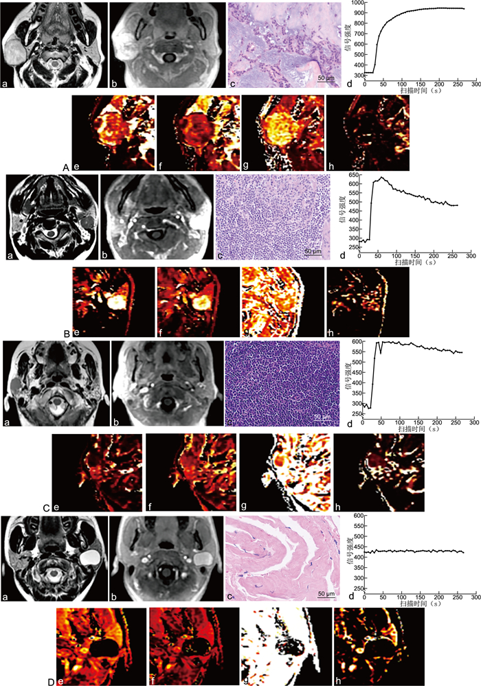
| Citation: | WANG Yan, QU Yuan, CHEN Jie, TIAN Hui, SHANG Yurong. Value of dynamic enhanced imaging radiomics in differentiating benign and malignant parotid gland neoplastic lesions[J]. Chinese Journal of General Practice, 2023, 21(3): 463-468. doi: 10.16766/j.cnki.issn.1674-4152.002908

|

| [1] |
唐晨虎, 马东, 孙伟, 等. 1.5T MR影像特征与腮腺肿瘤病人良恶性诊断的关系及病理结果对比[J]. 分子影像学杂志, 2021, 44(1): 127-131. https://www.cnki.com.cn/Article/CJFDTOTAL-FZYX202101025.htm
TANG C H, MA D, SUN W, et al. Relationship between 1.5 T MR imaging features and the diagnosis of parotid tumor patients[J]. Journal of Molecular Imaging, 2021, 44(1): 127-131. https://www.cnki.com.cn/Article/CJFDTOTAL-FZYX202101025.htm
|
| [2] |
ABDEL RAZEK A A K, MUKHERJI S K. State-of-the-art imaging of salivary gland tumors[J]. Neuroimaging Clin N Am, 2018, 28(2): 303-317. doi: 10.1016/j.nic.2018.01.009
|
| [3] |
COUDERT H, MIRAFZAL S, DISSARD A, et al. Multiparametric magnetic resonance imaging of parotid tumors: a systematic review[J]. Diagn Interv Imaging, 2021, 102(3): 121-130. doi: 10.1016/j.diii.2020.08.002
|
| [4] |
王海滨, 王理, 张乐星. 动态增强磁共振成像在腮腺肿瘤的诊断优势及研究进展[J]. 肿瘤学杂志, 2018, 24(6): 601-605. doi: 10.3760/cma.j.issn.1004-4221.2018.06.014
WANG H B, WANG L, ZHANG L X, et al. Research progress on diagnostic advantages of dynamic contrast-enhanced magnetic resonance imaging in parotid gland tumors[J]. Journal of Chinese Oncology, 2018, 24(6): 601-605. doi: 10.3760/cma.j.issn.1004-4221.2018.06.014
|
| [5] |
XU Z F, ZHENG S Y, PAN A Z, et al. A multiparametric analysis based on DCE-MRI to improve the accuracy of parotid tumor discrimination[J]. Eur J Nucl Med Mol Imaging, 2019, 46(11): 2228-2234. doi: 10.1007/s00259-019-04447-9
|
| [6] |
NADA A, HADY D, YOUSSEF A, et al. Accuracy of combined quantitative diffusion-weighted MRI and routine contrast-enhanced MRI in discrimination of benign and malignant salivary gland tumors[J]. Neuroradiol J, 2020, 33(3): 216-223. doi: 10.1177/1971400920913973
|
| [7] |
俞顺, 石清磊, 苏家威, 等. DCE-MRI定量参数在不同病理类型腮腺肿瘤鉴别诊断中的初步研究[J]. 磁共振成像, 2017, 8(9): 654-661. https://www.cnki.com.cn/Article/CJFDTOTAL-CGZC201709005.htm
YU S, SHI Q L, SU J W, et al. An initial study using the quantitative parameters of dynamic contrast-enhanced MRI in differential diagnosis for parotid tumors with different pathological types[J]. Chinese Journal of Magnetic Resonance Imaging, 2017, 8(9): 654-661. https://www.cnki.com.cn/Article/CJFDTOTAL-CGZC201709005.htm
|
| [8] |
ZHENG Y M, LI J, LIU S, et al. MRI-Based radiomics nomogram for differentiation of benign and malignant lesions of the parotid gland[J]. Eur Radiol, 2021, 31(6): 4042-4052. doi: 10.1007/s00330-020-07483-4
|
| [9] |
PILUDU F, MARZI S, RAVANELLI M, et al. MRI-based radiomics to differentiate between benign and malignant parotid tumors with external validation[J]. Front Oncol, 2021, 11: 656918. DOI: 10.3389/fonc.2021.656918.
|
| [10] |
吴艳, 谢元亮, 张树桐, 等. 基于T2WI影像组学及联合诊断模型鉴别腮腺多形性腺瘤与腺淋巴瘤[J]. 放射学实践, 2020, 35(12): 1525-1531. https://www.cnki.com.cn/Article/CJFDTOTAL-FSXS202012008.htm
WU Y, XIE Y L, ZHANG S T, et al. Radiomic analysis based on MR-T2WI and combined diagnostic model for identification of pleomorphic adenoma from adenolymphoma of parotid gland[J]. Radiologic Practice, 2020, 35(12): 1525-1531. https://www.cnki.com.cn/Article/CJFDTOTAL-FSXS202012008.htm
|
| [11] |
GÜNDÜZ E, ALÇIN Ö F, KIZILAY A, et al. Radiomics and deep learning approach to the differential diagnosis of parotid gland tumors[J]. Curr Opin Otolaryngol Head Neck Surg, 2022, 30(2): 107-113. doi: 10.1097/MOO.0000000000000782
|
| [12] |
DEBUS C, FLOCA R, INGRISCH M, et al. MITK-ModelFit: a generic open-source framework for model fits and their exploration in medical imaging-design, implementation and application on the example of DCE-MRI[J]. BMC Bioinformatics, 2019, 20(1): 31. doi: 10.1186/s12859-018-2588-1
|
| [13] |
SONG Y, ZHANG J, ZHANG Y D, et al. FeAture Explorer (FAE): a tool for developing and comparing radiomics models[J]. PLoS One, 2020, 15(8): e0237587. DOI: 10.1371/journal.pone.0237587.
|
| [14] |
江晓勇, 杨志辉, 陈希希, 等. 不同类型腮腺肿瘤的临床特点及远期复发情况分析[J]. 中华全科医学, 2019, 17(5): 790-792, 796. doi: 10.16766/j.cnki.issn.1674-4152.000793
JIANG X Y, YANG Z H, CHEN X X, et al. Analysis of clinical characteristics and long-term recurrence of different types of parotid tumors[J]. Chinese Journal of General Practice, 2019, 17(5): 790-792, 796. doi: 10.16766/j.cnki.issn.1674-4152.000793
|
| [15] |
GÖKÇE E, BEYHAN M. Advanced magnetic resonance imaging findings in salivary gland tumors[J]. World J Radiol, 2022, 14(8): 256-271. doi: 10.4329/wjr.v14.i8.256
|
| [16] |
胡涛, 刘琼, 邹玉坚, 等. 扩散峰度成像及动态增强MRI鉴别腮腺多形性腺瘤与Warthin瘤[J]. 放射学实践, 2021, 36(9): 1089-1094. https://www.cnki.com.cn/Article/CJFDTOTAL-FSXS202109009.htm
HU T, LIU Q, ZOU Y J, et al. Application value of DKI and DCE-MRI in the differential diagnosis of parotid pleomorphic adenoma and Warthin tumor[J]. Radiologic Practice, 2021, 36(9): 1089-1094. https://www.cnki.com.cn/Article/CJFDTOTAL-FSXS202109009.htm
|
| [17] |
ZHENG N, LI R, LIU W J, et al. The diagnostic value of combining conventional, diffusion-weighted imaging and dynamic contrast-enhanced MRI for salivary gland tumors[J]. Br J Radiol, 2018, 91(1089): 20170707. DOI: 10.1259/bjr.20170707.
|
| [18] |
YABUUCHI H, KAMITANI T, SAGIYAMA K, et al. Characterization of parotid gland tumors: added value of permeability MR imaging to DWI and DCE-MRI[J]. Eur Radiol, 2020, 30(12): 6402-6412. doi: 10.1007/s00330-020-07004-3
|
| [19] |
CHANG Y J, HUANG T Y, LIU Y J, et al. Classification of parotid gland tumors by using multimodal MRI and deep learning[J]. NMR Biomed, 2021, 34(1): e4408. DOI: 10.1002/nbm.4408.
|
| [20] |
HUANG N, XIAO Z B, CHEN Y, et al. Quantitative dynamic contrast-enhanced MRI and readout segmentation of long variable echo-trains diffusion-weighted imaging in differentiating parotid gland tumors[J]. Neuroradiology, 2021, 63(10): 1709-1719. doi: 10.1007/s00234-021-02758-z
|
Figures(1) / Tables(4)
Address:No. 287, Changhuai Road, Bengbu, Anhui Province, 233004, P.R.ChinaphoneNo:0552-3066635; 0552-3051890Email:zhqkyx@163.com
Copyright © Chinese Journal of General Practice皖ICP备2020018345号-2
Supported by:
Beijing Renhe Information Technology Co., Ltd.


 DownLoad: